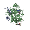
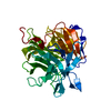
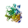
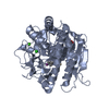
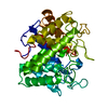
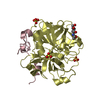
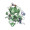
[English] 日本語
Yorodumi
- PDB-4az2: Human thrombin - inhibitor complex -

+
Open data


ID or keywords:

Loading...

-
Basic information

Entry
Database: PDB / ID: 4az2
TitleHuman thrombin - inhibitor complex
Components
  • HIRUDIN-3A'
  • THROMBIN HEAVY CHAIN
  • THROMBIN LIGHT CHAIN
KeywordsHYDROLASE/INHIBITOR / HYDROLASE-INHIBITOR COMPLEX
Function / homology
Function and homology information


positive regulation of lipid kinase activity / positive regulation of phospholipase C-activating G protein-coupled receptor signaling pathway / cytolysis by host of symbiont cells / thrombospondin receptor activity / Defective factor XII causes hereditary angioedema / thrombin / regulation of blood coagulation / neutrophil-mediated killing of gram-negative bacterium / ligand-gated ion channel signaling pathway / Defective F8 cleavage by thrombin ...positive regulation of lipid kinase activity / positive regulation of phospholipase C-activating G protein-coupled receptor signaling pathway / cytolysis by host of symbiont cells / thrombospondin receptor activity / Defective factor XII causes hereditary angioedema / thrombin / regulation of blood coagulation / neutrophil-mediated killing of gram-negative bacterium / ligand-gated ion channel signaling pathway / Defective F8 cleavage by thrombin / Platelet Aggregation (Plug Formation) / negative regulation of astrocyte differentiation / negative regulation of platelet activation / positive regulation of collagen biosynthetic process / negative regulation of cytokine production involved in inflammatory response / positive regulation of blood coagulation / negative regulation of fibrinolysis / Gamma-carboxylation of protein precursors / Transport of gamma-carboxylated protein precursors from the endoplasmic reticulum to the Golgi apparatus / Common Pathway of Fibrin Clot Formation / Removal of aminoterminal propeptides from gamma-carboxylated proteins / fibrinolysis / regulation of cytosolic calcium ion concentration / Intrinsic Pathway of Fibrin Clot Formation / Peptide ligand-binding receptors / positive regulation of release of sequestered calcium ion into cytosol / acute-phase response / Regulation of Complement cascade / negative regulation of proteolysis / Cell surface interactions at the vascular wall / lipopolysaccharide binding / positive regulation of receptor signaling pathway via JAK-STAT / growth factor activity / serine-type endopeptidase inhibitor activity / positive regulation of insulin secretion / platelet activation / response to wounding / positive regulation of protein localization to nucleus / Golgi lumen / antimicrobial humoral immune response mediated by antimicrobial peptide / Regulation of Insulin-like Growth Factor (IGF) transport and uptake by Insulin-like Growth Factor Binding Proteins (IGFBPs) / positive regulation of reactive oxygen species metabolic process / blood coagulation / Thrombin signalling through proteinase activated receptors (PARs) / heparin binding / regulation of cell shape / positive regulation of cell growth / G alpha (q) signalling events / collagen-containing extracellular matrix / positive regulation of phosphatidylinositol 3-kinase/protein kinase B signal transduction / cell surface receptor signaling pathway / blood microparticle / positive regulation of protein phosphorylation / G protein-coupled receptor signaling pathway / endoplasmic reticulum lumen / serine-type endopeptidase activity / signaling receptor binding / calcium ion binding / positive regulation of cell population proliferation / proteolysis / extracellular space / extracellular exosome / extracellular region / plasma membrane
Similarity search - Function
Thrombin inhibitor hirudin / Hirudin / Proteinase inhibitor I14, hirudin / Hirudin/antistatin / Prothrombin/thrombin / Thrombin light chain / Thrombin light chain domain superfamily / Thrombin light chain / Kringle domain / Kringle ...Thrombin inhibitor hirudin / Hirudin / Proteinase inhibitor I14, hirudin / Hirudin/antistatin / Prothrombin/thrombin / Thrombin light chain / Thrombin light chain domain superfamily / Thrombin light chain / Kringle domain / Kringle / Kringle, conserved site / Kringle superfamily / Kringle domain signature. / Kringle domain profile. / Kringle domain / Vitamin K-dependent carboxylation/gamma-carboxyglutamic (GLA) domain / Gamma-carboxyglutamic acid-rich (GLA) domain / Gamma-carboxyglutamic acid-rich (GLA) domain superfamily / Vitamin K-dependent carboxylation domain. / Gla domain profile. / Domain containing Gla (gamma-carboxyglutamate) residues. / Kringle-like fold / Serine proteases, trypsin family, histidine active site / Serine proteases, trypsin family, serine active site / Peptidase S1A, chymotrypsin family / Serine proteases, trypsin family, histidine active site. / Serine proteases, trypsin domain profile. / Serine proteases, trypsin family, serine active site. / Trypsin-like serine protease / Serine proteases, trypsin domain / Trypsin / Trypsin-like serine proteases / Thrombin, subunit H / Peptidase S1, PA clan, chymotrypsin-like fold / Peptidase S1, PA clan / Beta Barrel / Mainly Beta
Similarity search - Domain/homology
Chem-9MU / Prothrombin / Hirudin-3A' / Hirudin-3
Similarity search - Component
Biological speciesHOMO SAPIENS (human)
HIRUDO MEDICINALIS (medicinal leech)
MethodX-RAY DIFFRACTION / MOLECULAR REPLACEMENT / Resolution: 2.6 Å
AuthorsBanner, D.W. / D'Arcy, A. / Winkler, F.K. / Hilpert, K.
CitationJournal: J.Med.Chem. / Year: 1994
Title: Design and Synthesis of Potent and Highly Selective Thrombin Inhibitors.
Authors: Hilpert, K. / Ackermann, J. / Banner, D.W. / Gast, A. / Gubernator, K. / Hadvary, P. / Labler, L. / Muller, K. / Schmid, G. / Tschopp, T.B.
History
DepositionJun 22, 2012Deposition site: PDBE / Processing site: PDBE
Revision 1.0Aug 15, 2012Provider: repository / Type: Initial release
Revision 1.1Aug 7, 2013Group: Other
Revision 1.2Jun 28, 2017Group: Data collection / Category: diffrn_source
Item: _diffrn_source.pdbx_wavelength_list / _diffrn_source.type
Revision 1.3Jul 29, 2020Group: Data collection / Derived calculations ...Data collection / Derived calculations / Other / Structure summary
Category: chem_comp / entity ...chem_comp / entity / pdbx_chem_comp_identifier / pdbx_database_status / pdbx_entity_nonpoly / struct_conn / struct_site / struct_site_gen
Item: _chem_comp.name / _chem_comp.type ..._chem_comp.name / _chem_comp.type / _entity.pdbx_description / _pdbx_database_status.status_code_sf / _pdbx_entity_nonpoly.name / _struct_conn.pdbx_leaving_atom_flag / _struct_conn.pdbx_role
Description: Carbohydrate remediation / Provider: repository / Type: Remediation
Revision 1.4May 1, 2024Group: Data collection / Database references ...Data collection / Database references / Refinement description / Structure summary
Category: chem_comp / chem_comp_atom ...chem_comp / chem_comp_atom / chem_comp_bond / database_2 / pdbx_initial_refinement_model
Item: _chem_comp.pdbx_synonyms / _database_2.pdbx_DOI / _database_2.pdbx_database_accession
Remark 700 SHEET DETERMINATION METHOD: DSSP THE SHEETS PRESENTED AS "BB" IN EACH CHAIN ON SHEET RECORDS BELOW ... SHEET DETERMINATION METHOD: DSSP THE SHEETS PRESENTED AS "BB" IN EACH CHAIN ON SHEET RECORDS BELOW IS ACTUALLY AN 6-STRANDED BARREL THIS IS REPRESENTED BY A 7-STRANDED SHEET IN WHICH THE FIRST AND LAST STRANDS ARE IDENTICAL.

-
Structure visualization

Structure viewerMolecule:
MolmilJmol/JSmol

Downloads & links

-
Assembly

Deposited unit
A: THROMBIN LIGHT CHAIN
B: THROMBIN HEAVY CHAIN
D: HIRUDIN-3A'
hetero molecules


Theoretical massNumber of molelcules
Total (without water)35,2105
Polymers34,4953
Non-polymers7152
Water1,36976
1


  • Idetical with deposited unit
  • defined by author&software
TypeNameSymmetry operationNumber
identity operation1_555x,y,z1
Buried area3520 Å2
ΔGint-14.8 kcal/mol
Surface area14480 Å2
MethodPISA
Unit cell
Length a, b, c (Å)90.800, 90.800, 132.500
Angle α, β, γ (deg.)90.00, 90.00, 90.00
Int Tables number96
Space group name H-MP43212

-
Components

-
Protein/peptide , 2 types, 2 molecules AD

#1: Protein/peptide THROMBIN LIGHT CHAIN / COAGULATION FACTOR II


Mass: 3489.881 Da / Num. of mol.: 1 / Fragment: LIGHT CHAIN, RESIDUES 332-361 / Source method: isolated from a natural source / Source: (natural) HOMO SAPIENS (human) / References: UniProt: P00734, thrombin
#3: Protein/peptide HIRUDIN-3A' / HIRUDIN / HIRUDIN IIIA'


Mass: 1411.465 Da / Num. of mol.: 1 / Fragment: C-TERMINUS, RESIDUES 55-65 / Source method: obtained synthetically / Source: (synth.) HIRUDO MEDICINALIS (medicinal leech) / References: UniProt: P28509, UniProt: P28508*PLUS

-
Protein / Sugars , 2 types, 2 molecules B

#2: Protein THROMBIN HEAVY CHAIN / COAGULATION FACTOR II


Mass: 29594.055 Da / Num. of mol.: 1 / Fragment: HEAVY CHAIN, RESIDUES 364-620 / Source method: isolated from a natural source / Details: ASN B53 IS GLYCOSYLATED / Source: (natural) HOMO SAPIENS (human) / References: UniProt: P00734, thrombin
#5: Sugar ChemComp-NAG / 2-acetamido-2-deoxy-beta-D-glucopyranose / N-acetyl-beta-D-glucosamine / 2-acetamido-2-deoxy-beta-D-glucose / 2-acetamido-2-deoxy-D-glucose / 2-acetamido-2-deoxy-glucose / N-ACETYL-D-GLUCOSAMINE


Type: D-saccharide, beta linking / Mass: 221.208 Da / Num. of mol.: 1
Source method: isolated from a genetically manipulated source
Formula: C8H15NO6
IdentifierTypeProgram
DGlcpNAcbCONDENSED IUPAC CARBOHYDRATE SYMBOLGMML 1.0
N-acetyl-b-D-glucopyranosamineCOMMON NAMEGMML 1.0
b-D-GlcpNAcIUPAC CARBOHYDRATE SYMBOLPDB-CARE 1.0
GlcNAcSNFG CARBOHYDRATE SYMBOLGMML 1.0

-
Non-polymers , 2 types, 77 molecules

#4: Chemical ChemComp-9MU / (R)-N-((S)-1-CARBAMIMIDOYL-PIPERIDIN-3-YLMETHYL)-2-(NAPHTHALENE-2-SULFONYLAMINO)-3-PHENYL-PROPIONAMIDE


Mass: 493.621 Da / Num. of mol.: 1 / Source method: obtained synthetically / Formula: C26H31N5O3S
#6: Water ChemComp-HOH / water


Mass: 18.015 Da / Num. of mol.: 76 / Source method: isolated from a natural source / Formula: H2O

-
Details

Sequence detailsTHE N-TERMINAL RESIDUE IS MODIFIED IN CHAIN D. PLEASE REFER TO THE CITATION FOR FURTHER DETAILS.

-
Experimental details

-
Experiment

ExperimentMethod: X-RAY DIFFRACTION / Number of used crystals: 1

-
Sample preparation

CrystalDensity Matthews: 4.15 Å3/Da / Density % sol: 70.4 % / Description: SOME DATA PROCESSING INFORMATION HAS BEEN LOST

-
Data collection

DiffractionMean temperature: 15 K
Diffraction sourceSource: ROTATING ANODE / Type: ELLIOTT GX-21 / Wavelength: 1.5418 / Wavelength: 1.5418 Å
DetectorType: MULTIWIRE XENTRONICS / Detector: AREA DETECTOR / Date: Jan 6, 1994
RadiationMonochromator: NI FILTER / Protocol: SINGLE WAVELENGTH / Monochromatic (M) / Laue (L): M / Scattering type: x-ray
Radiation wavelengthWavelength: 1.5418 Å / Relative weight: 1
ReflectionResolution: 2.6→50 Å / Num. obs: 16462 / Observed criterion σ(I): 0 / Redundancy: 2.83 % / Rmerge(I) obs: 0.05
Reflection shellResolution: 2.6→2.7 Å / % possible all: 65

-
Processing

Software
NameVersionClassification
REFMAC5.7.0025refinement
XDSdata reduction
XDSdata scaling
AMoREphasing
RefinementMethod to determine structure: MOLECULAR REPLACEMENT
Starting model: IN HOUSE STRUCTURES

Resolution: 2.6→17.98 Å / Cor.coef. Fo:Fc: 0.967 / Cor.coef. Fo:Fc free: 0.943 / SU B: 5.893 / SU ML: 0.126 / Cross valid method: THROUGHOUT / ESU R: 0.276 / ESU R Free: 0.213 / Stereochemistry target values: MAXIMUM LIKELIHOOD
Details: HYDROGENS GENERATED AT RIDING POSITIONS IN REFMAC BUT NOT OUTPUT. RESIDUES B153 AND B154 HAVE ELECTRON DENSITY WHICH IS HARD TO FIT WITH THE PUBLISHED THROMBIN SEQUENCE.
RfactorNum. reflection% reflectionSelection details
Rfree0.19004 827 5.2 %RANDOM
Rwork0.14085 ---
obs0.14329 15223 90.85 %-
Solvent computationIon probe radii: 0.8 Å / Shrinkage radii: 0.8 Å / VDW probe radii: 1.2 Å / Solvent model: BABINET MODEL WITH MASK
Displacement parametersBiso mean: 38.412 Å2
Baniso -1Baniso -2Baniso -3
1-0.12 Å20 Å20 Å2
2--0.12 Å20 Å2
3----0.25 Å2
Refinement stepCycle: LAST / Resolution: 2.6→17.98 Å
ProteinNucleic acidLigandSolventTotal
Num. atoms2424 0 49 76 2549
Refine LS restraints
Refine-IDTypeDev idealDev ideal targetNumber
X-RAY DIFFRACTIONr_bond_refined_d0.0110.022552
X-RAY DIFFRACTIONr_bond_other_d
X-RAY DIFFRACTIONr_angle_refined_deg1.5271.9823453
X-RAY DIFFRACTIONr_angle_other_deg
X-RAY DIFFRACTIONr_dihedral_angle_1_deg6.6255297
X-RAY DIFFRACTIONr_dihedral_angle_2_deg36.74423.525122
X-RAY DIFFRACTIONr_dihedral_angle_3_deg15.80715446
X-RAY DIFFRACTIONr_dihedral_angle_4_deg14.3311521
X-RAY DIFFRACTIONr_chiral_restr0.1010.2355
X-RAY DIFFRACTIONr_gen_planes_refined0.0060.0212030
X-RAY DIFFRACTIONr_gen_planes_other
X-RAY DIFFRACTIONr_nbd_refined
X-RAY DIFFRACTIONr_nbd_other
X-RAY DIFFRACTIONr_nbtor_refined
X-RAY DIFFRACTIONr_nbtor_other
X-RAY DIFFRACTIONr_xyhbond_nbd_refined
X-RAY DIFFRACTIONr_xyhbond_nbd_other
X-RAY DIFFRACTIONr_metal_ion_refined
X-RAY DIFFRACTIONr_metal_ion_other
X-RAY DIFFRACTIONr_symmetry_vdw_refined
X-RAY DIFFRACTIONr_symmetry_vdw_other
X-RAY DIFFRACTIONr_symmetry_hbond_refined
X-RAY DIFFRACTIONr_symmetry_hbond_other
X-RAY DIFFRACTIONr_symmetry_metal_ion_refined
X-RAY DIFFRACTIONr_symmetry_metal_ion_other
X-RAY DIFFRACTIONr_mcbond_it
X-RAY DIFFRACTIONr_mcbond_other
X-RAY DIFFRACTIONr_mcangle_it
X-RAY DIFFRACTIONr_mcangle_other
X-RAY DIFFRACTIONr_scbond_it
X-RAY DIFFRACTIONr_scbond_other
X-RAY DIFFRACTIONr_scangle_it
X-RAY DIFFRACTIONr_scangle_other
X-RAY DIFFRACTIONr_long_range_B_refined
X-RAY DIFFRACTIONr_long_range_B_other
X-RAY DIFFRACTIONr_rigid_bond_restr
X-RAY DIFFRACTIONr_sphericity_free
X-RAY DIFFRACTIONr_sphericity_bonded
LS refinement shellResolution: 2.601→2.668 Å / Total num. of bins used: 20
RfactorNum. reflection% reflection
Rfree0.302 36 -
Rwork0.184 651 -
obs--53.71 %

+
About Yorodumi

-
News

-
Feb 9, 2022. New format data for meta-information of EMDB entries

New format data for meta-information of EMDB entries

  • Version 3 of the EMDB header file is now the official format.
  • The previous official version 1.9 will be removed from the archive.

Related info.:EMDB header

External links:wwPDB to switch to version 3 of the EMDB data model

-
Aug 12, 2020. Covid-19 info

Covid-19 info

URL: https://pdbjlvh1.pdbj.org/emnavi/covid19.php

New page: Covid-19 featured information page in EM Navigator.

Related info.:Covid-19 info / Mar 5, 2020. Novel coronavirus structure data

+
Mar 5, 2020. Novel coronavirus structure data

Novel coronavirus structure data

Related info.:Yorodumi Speices / Aug 12, 2020. Covid-19 info

External links:COVID-19 featured content - PDBj / Molecule of the Month (242):Coronavirus Proteases

+
Jan 31, 2019. EMDB accession codes are about to change! (news from PDBe EMDB page)

EMDB accession codes are about to change! (news from PDBe EMDB page)

  • The allocation of 4 digits for EMDB accession codes will soon come to an end. Whilst these codes will remain in use, new EMDB accession codes will include an additional digit and will expand incrementally as the available range of codes is exhausted. The current 4-digit format prefixed with “EMD-” (i.e. EMD-XXXX) will advance to a 5-digit format (i.e. EMD-XXXXX), and so on. It is currently estimated that the 4-digit codes will be depleted around Spring 2019, at which point the 5-digit format will come into force.
  • The EM Navigator/Yorodumi systems omit the EMD- prefix.

Related info.:Q: What is EMD? / ID/Accession-code notation in Yorodumi/EM Navigator

External links:EMDB Accession Codes are Changing Soon! / Contact to PDBj

+
Jul 12, 2017. Major update of PDB

Major update of PDB

  • wwPDB released updated PDB data conforming to the new PDBx/mmCIF dictionary.
  • This is a major update changing the version number from 4 to 5, and with Remediation, in which all the entries are updated.
  • In this update, many items about electron microscopy experimental information are reorganized (e.g. em_software).
  • Now, EM Navigator and Yorodumi are based on the updated data.

External links:wwPDB Remediation / Enriched Model Files Conforming to OneDep Data Standards Now Available in the PDB FTP Archive

-
Yorodumi

Thousand views of thousand structures

  • Yorodumi is a browser for structure data from EMDB, PDB, SASBDB, etc.
  • This page is also the successor to EM Navigator detail page, and also detail information page/front-end page for Omokage search.
  • The word "yorodu" (or yorozu) is an old Japanese word meaning "ten thousand". "mi" (miru) is to see.

Related info.:EMDB / PDB / SASBDB / Comparison of 3 databanks / Yorodumi Search / Aug 31, 2016. New EM Navigator & Yorodumi / Yorodumi Papers / Jmol/JSmol / Function and homology information / Changes in new EM Navigator and Yorodumi

Read more