 Movie
Movie Controller

[English] 日本語
 Yorodumi
- PDB-4ayp: Structure of The GH47 processing alpha-1,2-mannosidase from Caulo... -
+ Open data

- Basic information
| Entry | Database: PDB / ID: 4ayp | |||||||||
|---|---|---|---|---|---|---|---|---|---|---|
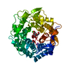
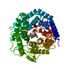
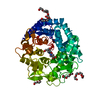
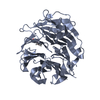
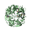
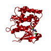
| Title | Structure of The GH47 processing alpha-1,2-mannosidase from Caulobacter strain K31 in complex with thiomannobioside | |||||||||
 Components Components | MANNOSYL-OLIGOSACCHARIDE 1,2-ALPHA-MANNOSIDASE | |||||||||
 Keywords Keywords | HYDROLASE / GLYCOSIDE HYDROLASE / GH47 / CAZY / ENZYME-CARBOHYDRATE INTERACTION / MANNOSE / GLYCOSIDASE INHIBITION / QUANTUM MECHANICS | |||||||||
| Function / homology |  Function and homology information Function and homology informationmannosyl-oligosaccharide 1,2-alpha-mannosidase / mannosyl-oligosaccharide 1,2-alpha-mannosidase activity / endoplasmic reticulum mannose trimming / carbohydrate metabolic process / calcium ion binding / membrane Similarity search - Function | |||||||||
| Biological species |  CAULOBACTER SP. (bacteria) CAULOBACTER SP. (bacteria) | |||||||||
| Method |  X-RAY DIFFRACTION / SYNCHROTRON / MOLECULAR REPLACEMENT / Resolution: 0.85 Å X-RAY DIFFRACTION / SYNCHROTRON / MOLECULAR REPLACEMENT / Resolution: 0.85 Å | |||||||||
 Authors Authors | Thompson, A.J. / Dabin, J. / Iglesias-Fernandez, J. / Iglesias-Fernandez, A. / Dinev, Z. / Williams, S.J. / Siriwardena, A. / Moreland, C. / Hu, T.C. / Smith, D.K. ...Thompson, A.J. / Dabin, J. / Iglesias-Fernandez, J. / Iglesias-Fernandez, A. / Dinev, Z. / Williams, S.J. / Siriwardena, A. / Moreland, C. / Hu, T.C. / Smith, D.K. / Gilbert, H.J. / Rovira, C. / Davies, G.J. | |||||||||
 Citation Citation | Journal: Angew.Chem.Int.Ed.Engl. / Year: 2012 Title: The Reaction Coordinate of a Bacterial Gh47 Alpha-Mannosidase: A Combined Quantum Mechanical and Structural Approach. Authors: Thompson, A.J. / Dabin, J. / Iglesias-Fernandez, J. / Ardevol, A. / Dinev, Z. / Williams, S.J. / Bande, O. / Siriwardena, A. / Moreland, C. / Hu, T.C. / Smith, D.K. / Gilbert, H.J. / Rovira, C. / Davies, G.J. | |||||||||
| History |
|
- Structure visualization
| Structure viewer | Molecule: MolmilJmol/JSmol |
|---|
- Downloads & links
-Download
| PDBx/mmCIF format | 4ayp.cif.gz | 224.2 KB | Display | PDBx/mmCIF format |
|---|---|---|---|---|
| PDB format | pdb4ayp.ent.gz | 177.5 KB | Display | PDB format |
| PDBx/mmJSON format | 4ayp.json.gz | Tree view | PDBx/mmJSON format | |
| Others |  Other downloads Other downloads |
-Validation report
| Arichive directory | https://data.pdbj.org/pub/pdb/validation_reports/ay/4aypftp://data.pdbj.org/pub/pdb/validation_reports/ay/4ayp | HTTPS FTP |
|---|
-Related structure data


-Links
 PDBj
PDBj


- Assembly
| Deposited unit | 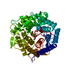
| ||||||||
|---|---|---|---|---|---|---|---|---|---|
| 1 |
| ||||||||
| Unit cell |
|
-Components
| #1: Protein | Mass: 50546.676 Da / Num. of mol.: 1 Source method: isolated from a genetically manipulated source Source: (gene. exp.) CAULOBACTER SP. (bacteria) / Strain: K31 / Production host: References: UniProt: B0SWV2, mannosyl-oligosaccharide 1,2-alpha-mannosidase | ||||||
|---|---|---|---|---|---|---|---|
| #2: Polysaccharide | alpha-D-mannopyranose-(1-2)-methyl 2-thio-alpha-D-mannopyranoside / methyl 2-S-alpha-D-mannopyranosyl-2-thio-alpha-D-mannopyranoside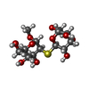  Type: oligosaccharide, Oligosaccharide / Class: Substrate analog / Mass: 372.389 Da / Num. of mol.: 1 Type: oligosaccharide, Oligosaccharide / Class: Substrate analog / Mass: 372.389 Da / Num. of mol.: 1Source method: isolated from a genetically manipulated source Details: oligosaccharide with S-glycosidic bond between monosaccharides References: methyl 2-S-alpha-D-mannopyranosyl-2-thio-alpha-D-mannopyranoside | ||||||
| #3: Chemical |   Mass: 40.078 Da / Num. of mol.: 2 / Source method: obtained synthetically / Formula: Ca Mass: 40.078 Da / Num. of mol.: 2 / Source method: obtained synthetically / Formula: Ca#4: Chemical | ChemComp-NA / |   Mass: 22.990 Da / Num. of mol.: 1 / Source method: obtained synthetically / Formula: Na Mass: 22.990 Da / Num. of mol.: 1 / Source method: obtained synthetically / Formula: Na#5: Water | ChemComp-HOH / |  Mass: 18.015 Da / Num. of mol.: 781 / Source method: isolated from a natural source / Formula: H2O Mass: 18.015 Da / Num. of mol.: 781 / Source method: isolated from a natural source / Formula: H2OSequence details | N-TERMINAL TRUNCATION | |
-Experimental details
-Experiment
| Experiment | Method: X-RAY DIFFRACTION / Number of used crystals: 1 |
|---|
- Sample preparation
| Crystal | Density Matthews: 2 Å3/Da / Density % sol: 38.6 % / Description: NONE |
|---|---|
| Crystal grow | pH: 6.5 Details: 0.2 M AMMONIUM ACTETATE, 0.1 M BIS-TRIS PH 6.5, 22% WT/VOL PEG 3350 |
-Data collection
| Diffraction | Mean temperature: 100 K |
|---|---|
| Diffraction source | Source: SYNCHROTRON / Site: Diamond  / Beamline: I03 / Wavelength: 0.7749 / Beamline: I03 / Wavelength: 0.7749 |
| Detector | Type: DECTRIS PILATUS 6M / Detector: PIXEL / Date: Jul 11, 2011 / Details: MIRRORS |
| Radiation | Protocol: SINGLE WAVELENGTH / Monochromatic (M) / Laue (L): M / Scattering type: x-ray |
| Radiation wavelength | Wavelength: 0.7749 Å / Relative weight: 1 |
| Reflection | Resolution: 0.85→46.65 Å / Num. obs: 333281 / % possible obs: 97.2 % / Observed criterion σ(I): 1.7 / Redundancy: 5.1 % / Rmerge(I) obs: 0.04 / Net I/σ(I): 16.9 |
| Reflection shell | Resolution: 0.85→0.9 Å / Redundancy: 2.8 % / Rmerge(I) obs: 0.57 / Mean I/σ(I) obs: 1.7 / % possible all: 80.8 |
- Processing
| Software |
| ||||||||||||||||||||||||||||||||||||||||||||||||||||||||||||||||||||||||||||||||||||||||||||||||||||||||||||||||||||||||||||||||||||||||||||||||||||||||||||||||||||||||||||||||||||||
|---|---|---|---|---|---|---|---|---|---|---|---|---|---|---|---|---|---|---|---|---|---|---|---|---|---|---|---|---|---|---|---|---|---|---|---|---|---|---|---|---|---|---|---|---|---|---|---|---|---|---|---|---|---|---|---|---|---|---|---|---|---|---|---|---|---|---|---|---|---|---|---|---|---|---|---|---|---|---|---|---|---|---|---|---|---|---|---|---|---|---|---|---|---|---|---|---|---|---|---|---|---|---|---|---|---|---|---|---|---|---|---|---|---|---|---|---|---|---|---|---|---|---|---|---|---|---|---|---|---|---|---|---|---|---|---|---|---|---|---|---|---|---|---|---|---|---|---|---|---|---|---|---|---|---|---|---|---|---|---|---|---|---|---|---|---|---|---|---|---|---|---|---|---|---|---|---|---|---|---|---|---|---|---|
| Refinement | Method to determine structure: MOLECULAR REPLACEMENT Starting model: PREVIOUSLY SOLVED NATIVE STRUCTURE Resolution: 0.85→39.16 Å / Cor.coef. Fo:Fc: 0.99 / Cor.coef. Fo:Fc free: 0.987 / SU B: 0.232 / SU ML: 0.006 / Cross valid method: THROUGHOUT / ESU R: 0.011 / ESU R Free: 0.011 / Stereochemistry target values: MAXIMUM LIKELIHOOD Details: HYDROGENS HAVE BEEN ADDED IN THE RIDING POSITIONS. U VALUES REFINED INDIVIDUALLY
| ||||||||||||||||||||||||||||||||||||||||||||||||||||||||||||||||||||||||||||||||||||||||||||||||||||||||||||||||||||||||||||||||||||||||||||||||||||||||||||||||||||||||||||||||||||||
| Solvent computation | Ion probe radii: 0.8 Å / Shrinkage radii: 0.8 Å / VDW probe radii: 1.2 Å / Solvent model: MASK | ||||||||||||||||||||||||||||||||||||||||||||||||||||||||||||||||||||||||||||||||||||||||||||||||||||||||||||||||||||||||||||||||||||||||||||||||||||||||||||||||||||||||||||||||||||||
| Displacement parameters | Biso mean: 8.802 Å2
| ||||||||||||||||||||||||||||||||||||||||||||||||||||||||||||||||||||||||||||||||||||||||||||||||||||||||||||||||||||||||||||||||||||||||||||||||||||||||||||||||||||||||||||||||||||||
| Refinement step | Cycle: LAST / Resolution: 0.85→39.16 Å
| ||||||||||||||||||||||||||||||||||||||||||||||||||||||||||||||||||||||||||||||||||||||||||||||||||||||||||||||||||||||||||||||||||||||||||||||||||||||||||||||||||||||||||||||||||||||
| Refine LS restraints |
|