Mass: 18.015 Da / Num. of mol.: 326 / Source method: isolated from a natural source / Formula: H2O
-
Details
Has protein modification
Y
Nonpolymer details
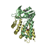
STATINE (STA): PART OF PEPSTATIN A ISOVALERIC ACID (IVA): PART OF PEPSTATIN A
-
Experimental details
-
Experiment
Experiment
Method: X-RAY DIFFRACTION / Number of used crystals: 1
-
Sample preparation
Crystal
Density Matthews: 3.1 Å3/Da / Density % sol: 60 % / Description: NONE
Crystal grow
pH: 5.5 Details: RESERVOIR: 75 MM BIS-TRIS PH 5.5, 0.15 M NACL, 19% (W/V) PEG3350, 7.5 MM CDCL2. SAMPLE: 7.2 MG/ML OF PROTEIN COMPLEX IN 10 MM NAH2PO4 PH 6
Resolution: 1.6→41.388 Å / SU ML: 0.36 / σ(F): 2.01 / Phase error: 17.44 / Stereochemistry target values: ML Details: DISORDERED LOOP 159-160 WAS MODELED STEREOCHEMICALLY. VM CALCULATED USING A TOTAL MASS OF 36.3 KDA CORRESPONDING CHYMOSIN INHIBITED BY PEPSTATIN
Rfactor
Num. reflection
% reflection
Rfree
0.1971
3009
5.1 %
Rwork
0.1743
-
-
obs
0.1754
59607
99.94 %
Solvent computation
Shrinkage radii: 0.73 Å / VDW probe radii: 1 Å / Solvent model: FLAT BULK SOLVENT MODEL / Bsol: 41.504 Å2 / ksol: 0.36 e/Å3
Displacement parameters
Biso mean: 24.7 Å2
Baniso -1
Baniso -2
Baniso -3
1-
-1.0212 Å2
0 Å2
0 Å2
2-
-
-1.0212 Å2
0 Å2
3-
-
-
2.0424 Å2
Refinement step
Cycle: LAST / Resolution: 1.6→41.388 Å
Protein
Nucleic acid
Ligand
Solvent
Total
Num. atoms
2559
0
18
326
2903
Refine LS restraints
Refine-ID
Type
Dev ideal
Number
X-RAY DIFFRACTION
f_bond_d
0.019
2689
X-RAY DIFFRACTION
f_angle_d
1.249
3651
X-RAY DIFFRACTION
f_dihedral_angle_d
14.365
957
X-RAY DIFFRACTION
f_chiral_restr
0.095
406
X-RAY DIFFRACTION
f_plane_restr
0.006
473
LS refinement shell
Resolution (Å)
Rfactor Rfree
Num. reflection Rfree
Rfactor Rwork
Num. reflection Rwork
Refine-ID
% reflection obs (%)
1.6-1.6262
0.2216
134
0.2173
2675
X-RAY DIFFRACTION
100
1.6262-1.6543
0.2432
136
0.2229
2700
X-RAY DIFFRACTION
100
1.6543-1.6844
0.2365
156
0.2183
2641
X-RAY DIFFRACTION
100
1.6844-1.7168
0.2635
144
0.2103
2653
X-RAY DIFFRACTION
100
1.7168-1.7518
0.2168
135
0.2009
2669
X-RAY DIFFRACTION
100
1.7518-1.7899
0.2066
131
0.2016
2683
X-RAY DIFFRACTION
100
1.7899-1.8315
0.2174
158
0.1916
2646
X-RAY DIFFRACTION
100
1.8315-1.8773
0.2115
154
0.182
2670
X-RAY DIFFRACTION
100
1.8773-1.9281
0.2123
126
0.1779
2707
X-RAY DIFFRACTION
100
1.9281-1.9848
0.1871
151
0.1781
2657
X-RAY DIFFRACTION
100
1.9848-2.0489
0.1909
143
0.1779
2684
X-RAY DIFFRACTION
100
2.0489-2.1221
0.2038
139
0.1803
2694
X-RAY DIFFRACTION
100
2.1221-2.2071
0.2233
144
0.1798
2669
X-RAY DIFFRACTION
100
2.2071-2.3075
0.2157
164
0.1802
2680
X-RAY DIFFRACTION
100
2.3075-2.4292
0.202
148
0.1792
2690
X-RAY DIFFRACTION
100
2.4292-2.5813
0.1906
122
0.1832
2717
X-RAY DIFFRACTION
100
2.5813-2.7806
0.2161
152
0.1803
2685
X-RAY DIFFRACTION
100
2.7806-3.0603
0.2122
142
0.1729
2725
X-RAY DIFFRACTION
100
3.0603-3.503
0.1831
145
0.1628
2730
X-RAY DIFFRACTION
100
3.503-4.4126
0.1484
153
0.1444
2745
X-RAY DIFFRACTION
100
4.4126-41.4014
0.2006
132
0.1694
2878
X-RAY DIFFRACTION
100
+
About Yorodumi
-
News
-
Feb 9, 2022. New format data for meta-information of EMDB entries
New format data for meta-information of EMDB entries
Version 3 of the EMDB header file is now the official format.
The previous official version 1.9 will be removed from the archive.
In the structure databanks used in Yorodumi, some data are registered as the other names, "COVID-19 virus" and "2019-nCoV". Here are the details of the virus and the list of structure data.
Jan 31, 2019. EMDB accession codes are about to change! (news from PDBe EMDB page)
EMDB accession codes are about to change! (news from PDBe EMDB page)
The allocation of 4 digits for EMDB accession codes will soon come to an end. Whilst these codes will remain in use, new EMDB accession codes will include an additional digit and will expand incrementally as the available range of codes is exhausted. The current 4-digit format prefixed with “EMD-” (i.e. EMD-XXXX) will advance to a 5-digit format (i.e. EMD-XXXXX), and so on. It is currently estimated that the 4-digit codes will be depleted around Spring 2019, at which point the 5-digit format will come into force.
The EM Navigator/Yorodumi systems omit the EMD- prefix.
Related info.:Q: What is EMD? / ID/Accession-code notation in Yorodumi/EM Navigator
Yorodumi is a browser for structure data from EMDB, PDB, SASBDB, etc.
This page is also the successor to EM Navigator detail page, and also detail information page/front-end page for Omokage search.
The word "yorodu" (or yorozu) is an old Japanese word meaning "ten thousand". "mi" (miru) is to see.
Related info.:EMDB / PDB / SASBDB / Comparison of 3 databanks / Yorodumi Search / Aug 31, 2016. New EM Navigator & Yorodumi / Yorodumi Papers / Jmol/JSmol / Function and homology information / Changes in new EM Navigator and Yorodumi
 Movie
Movie Controller


 Open data
 Basic information
 Components
 Keywords
 Function and homology information
 ACTINOMYCES (bacteria)
 X-RAY DIFFRACTION /
  Authors
 Citation
 Structure visualization
 Downloads & links
 Other downloads


 PDBj
PDBj
 Assembly


 Mass: 22.990 Da / Num. of mol.: 1 / Source method: obtained synthetically / Formula: Na
 Mass: 112.411 Da / Num. of mol.: 1 / Source method: obtained synthetically / Formula: Cd
 Mass: 62.068 Da / Num. of mol.: 4 / Source method: obtained synthetically / Formula: C2H6O2
 Sample preparation
 / Beamline: ID29 / Wavelength: 0.89993
  Processing