 Movie
Movie Controller

[English] 日本語
 Yorodumi
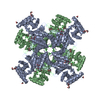
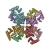
- PDB-4aub: the complex Structure of the bacterial aldo-keto reductase AKR14A... -
+ Open data

- Basic information
| Entry | Database: PDB / ID: 4aub | ||||||
|---|---|---|---|---|---|---|---|
| Title | the complex Structure of the bacterial aldo-keto reductase AKR14A1 with NADP and citrate | ||||||
 Components Components | ALDO-KETO REDUCTASE AKR14A1 | ||||||
 Keywords Keywords | OXIDOREDUCTASE | ||||||
| Function / homology |  Function and homology information Function and homology informationmethylglyoxal catabolic process / alcohol dehydrogenase (NADP+) / methylglyoxal reductase (NADPH) (acetol producing) activity / alcohol dehydrogenase (NADP+) activity / Oxidoreductases; Acting on the CH-OH group of donors; With NAD+ or NADP+ as acceptor / oxidoreductase activity, acting on the CH-OH group of donors, NAD or NADP as acceptor / DNA damage response Similarity search - Function | ||||||
| Biological species |  | ||||||
| Method |  X-RAY DIFFRACTION / MOLECULAR REPLACEMENT / Resolution: 2.05 Å X-RAY DIFFRACTION / MOLECULAR REPLACEMENT / Resolution: 2.05 Å | ||||||
 Authors Authors | Zhu, X. / Ellis, E.M. / Lapthorn, A. | ||||||
 Citation Citation | Journal: Chem.Biol.Interact / Year: 2013 Title: The Diversity of Microbial Aldo/Keto Reductases from Escherichia Coli K12. Authors: Lapthorn, A.J. / Zhu, X. / Ellis, E.M. | ||||||
| History |
| ||||||
| Remark 700 | SHEET DETERMINATION METHOD: DSSP THE SHEETS PRESENTED AS "AB" IN EACH CHAIN ON SHEET RECORDS BELOW ... SHEET DETERMINATION METHOD: DSSP THE SHEETS PRESENTED AS "AB" IN EACH CHAIN ON SHEET RECORDS BELOW IS ACTUALLY AN 8-STRANDED BARREL THIS IS REPRESENTED BY A 9-STRANDED SHEET IN WHICH THE FIRST AND LAST STRANDS ARE IDENTICAL. THE SHEETS PRESENTED AS "BB" IN EACH CHAIN ON SHEET RECORDS BELOW IS ACTUALLY AN 8-STRANDED BARREL THIS IS REPRESENTED BY A 9-STRANDED SHEET IN WHICH THE FIRST AND LAST STRANDS ARE IDENTICAL. THE SHEETS PRESENTED AS "CB" IN EACH CHAIN ON SHEET RECORDS BELOW IS ACTUALLY AN 8-STRANDED BARREL THIS IS REPRESENTED BY A 9-STRANDED SHEET IN WHICH THE FIRST AND LAST STRANDS ARE IDENTICAL. THE SHEETS PRESENTED AS "DB" IN EACH CHAIN ON SHEET RECORDS BELOW IS ACTUALLY AN 8-STRANDED BARREL THIS IS REPRESENTED BY A 9-STRANDED SHEET IN WHICH THE FIRST AND LAST STRANDS ARE IDENTICAL. THE SHEETS PRESENTED AS "EB" IN EACH CHAIN ON SHEET RECORDS BELOW IS ACTUALLY AN 8-STRANDED BARREL THIS IS REPRESENTED BY A 9-STRANDED SHEET IN WHICH THE FIRST AND LAST STRANDS ARE IDENTICAL. THE SHEETS PRESENTED AS "FB" IN EACH CHAIN ON SHEET RECORDS BELOW IS ACTUALLY AN 8-STRANDED BARREL THIS IS REPRESENTED BY A 9-STRANDED SHEET IN WHICH THE FIRST AND LAST STRANDS ARE IDENTICAL. THE SHEETS PRESENTED AS "GB" IN EACH CHAIN ON SHEET RECORDS BELOW IS ACTUALLY AN 8-STRANDED BARREL THIS IS REPRESENTED BY A 9-STRANDED SHEET IN WHICH THE FIRST AND LAST STRANDS ARE IDENTICAL. THE SHEETS PRESENTED AS "HB" IN EACH CHAIN ON SHEET RECORDS BELOW IS ACTUALLY AN 8-STRANDED BARREL THIS IS REPRESENTED BY A 9-STRANDED SHEET IN WHICH THE FIRST AND LAST STRANDS ARE IDENTICAL. |
- Structure visualization
| Structure viewer | Molecule: MolmilJmol/JSmol |
|---|
- Downloads & links
-Download
| PDBx/mmCIF format | 4aub.cif.gz | 526.9 KB | Display | PDBx/mmCIF format |
|---|---|---|---|---|
| PDB format | pdb4aub.ent.gz | 431.6 KB | Display | PDB format |
| PDBx/mmJSON format | 4aub.json.gz | Tree view | PDBx/mmJSON format | |
| Others |  Other downloads Other downloads |
-Validation report
| Arichive directory | https://data.pdbj.org/pub/pdb/validation_reports/au/4aubftp://data.pdbj.org/pub/pdb/validation_reports/au/4aub | HTTPS FTP |
|---|
-Related structure data
| Related structure data | 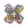 4astC 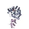 1exbS C: citing same article ( S: Starting model for refinement |
|---|---|
| Similar structure data |


-Links
 PDBj
PDBj


- Assembly
| Deposited unit | 
| ||||||||
|---|---|---|---|---|---|---|---|---|---|
| 1 |
| ||||||||
| Unit cell |
|
-Components
| #1: Protein | Mass: 41047.453 Da / Num. of mol.: 8 Source method: isolated from a genetically manipulated source Source: (gene. exp.) #2: Chemical | ChemComp-NAP /   Mass: 743.405 Da / Num. of mol.: 7 / Source method: obtained synthetically / Formula: C21H28N7O17P3 Mass: 743.405 Da / Num. of mol.: 7 / Source method: obtained synthetically / Formula: C21H28N7O17P3#3: Chemical | ChemComp-FLC /   Mass: 189.100 Da / Num. of mol.: 8 / Source method: obtained synthetically / Formula: C6H5O7 Mass: 189.100 Da / Num. of mol.: 8 / Source method: obtained synthetically / Formula: C6H5O7#4: Water | ChemComp-HOH / |  Mass: 18.015 Da / Num. of mol.: 1161 / Source method: isolated from a natural source / Formula: H2O Mass: 18.015 Da / Num. of mol.: 1161 / Source method: isolated from a natural source / Formula: H2O |
|---|
-Experimental details
-Experiment
| Experiment | Method: X-RAY DIFFRACTION / Number of used crystals: 1 |
|---|
- Sample preparation
| Crystal | Density Matthews: 2.68 Å3/Da / Density % sol: 54.2 % Description: STATISTICS ARE GOOD TO 2.21 ANGSTROM RESOLUTION, RMERGE 0.48 IN FINAL SHELL. HOWEVER THE WEAK REFLECTION EXTENDS TO 2.05 ANGSTROM RESOLUTION AND WAS INCLUDED IN REFINEMENT |
|---|---|
| Crystal grow | pH: 5.4 Details: 0.1M CITRATE BUFFER (PH 5.4), 18% W/V PEG 2000 MME AND 0.2 M AMMONIUM SULPHATE SUPPLEMENTED WITH 1 MM NADP |
-Data collection
| Diffraction | Mean temperature: 100 K |
|---|---|
| Diffraction source | Source: ROTATING ANODE / Wavelength: 1.5418 |
| Detector | Detector: CCD |
| Radiation | Protocol: SINGLE WAVELENGTH / Monochromatic (M) / Laue (L): M / Scattering type: x-ray |
| Radiation wavelength | Wavelength: 1.5418 Å / Relative weight: 1 |
| Reflection | Resolution: 2.05→48.8 Å / Num. obs: 194971 / % possible obs: 97.3 % / Observed criterion σ(I): 2 / Redundancy: 4.8 % / Rmerge(I) obs: 0.1 / Net I/σ(I): 20.3 |
| Reflection shell | Resolution: 2.05→2.09 Å / Redundancy: 4.6 % / Rmerge(I) obs: 0.87 / Mean I/σ(I) obs: 2.8 / % possible all: 95 |
- Processing
| Software |
| ||||||||||||||||||||||||||||||||||||||||||||||||||||||||||||||||||||||||||||||||||||||||||||||||||||||||||||||||||||||||||||||||||||||||||||||||||||||||||||||||||||||||||||||||||||||
|---|---|---|---|---|---|---|---|---|---|---|---|---|---|---|---|---|---|---|---|---|---|---|---|---|---|---|---|---|---|---|---|---|---|---|---|---|---|---|---|---|---|---|---|---|---|---|---|---|---|---|---|---|---|---|---|---|---|---|---|---|---|---|---|---|---|---|---|---|---|---|---|---|---|---|---|---|---|---|---|---|---|---|---|---|---|---|---|---|---|---|---|---|---|---|---|---|---|---|---|---|---|---|---|---|---|---|---|---|---|---|---|---|---|---|---|---|---|---|---|---|---|---|---|---|---|---|---|---|---|---|---|---|---|---|---|---|---|---|---|---|---|---|---|---|---|---|---|---|---|---|---|---|---|---|---|---|---|---|---|---|---|---|---|---|---|---|---|---|---|---|---|---|---|---|---|---|---|---|---|---|---|---|---|
| Refinement | Method to determine structure: MOLECULAR REPLACEMENT Starting model: PDB ENTRY 1EXB Resolution: 2.05→95.35 Å / Cor.coef. Fo:Fc: 0.936 / Cor.coef. Fo:Fc free: 0.914 / SU B: 4.57 / SU ML: 0.123 / Cross valid method: THROUGHOUT / ESU R: 0.211 / ESU R Free: 0.179 / Stereochemistry target values: MAXIMUM LIKELIHOOD / Details: HYDROGENS HAVE BEEN ADDED IN THE RIDING POSITIONS.
| ||||||||||||||||||||||||||||||||||||||||||||||||||||||||||||||||||||||||||||||||||||||||||||||||||||||||||||||||||||||||||||||||||||||||||||||||||||||||||||||||||||||||||||||||||||||
| Solvent computation | Ion probe radii: 0.8 Å / Shrinkage radii: 0.8 Å / VDW probe radii: 1.2 Å / Solvent model: MASK | ||||||||||||||||||||||||||||||||||||||||||||||||||||||||||||||||||||||||||||||||||||||||||||||||||||||||||||||||||||||||||||||||||||||||||||||||||||||||||||||||||||||||||||||||||||||
| Displacement parameters | Biso mean: 25.481 Å2
| ||||||||||||||||||||||||||||||||||||||||||||||||||||||||||||||||||||||||||||||||||||||||||||||||||||||||||||||||||||||||||||||||||||||||||||||||||||||||||||||||||||||||||||||||||||||
| Refinement step | Cycle: LAST / Resolution: 2.05→95.35 Å
| ||||||||||||||||||||||||||||||||||||||||||||||||||||||||||||||||||||||||||||||||||||||||||||||||||||||||||||||||||||||||||||||||||||||||||||||||||||||||||||||||||||||||||||||||||||||
| Refine LS restraints |
|