 Movie
Movie Controller

[English] 日本語
 Yorodumi
- PDB-4arh: X ray structure of the periplasmic zinc binding protein ZinT from... -
+ Open data

- Basic information
| Entry | Database: PDB / ID: 4arh | ||||||
|---|---|---|---|---|---|---|---|
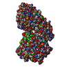
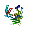
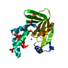
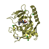
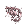
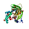
| Title | X ray structure of the periplasmic zinc binding protein ZinT from Salmonella enterica | ||||||
 Components Components | METAL-BINDING PROTEIN YODA | ||||||
 Keywords Keywords | METAL BINDING PROTEIN / PERIPLASMIC PROTEIN | ||||||
| Function / homology | Calycin beta-barrel core domain / Lipocalin / Beta Barrel / Mainly Beta / :  Function and homology information Function and homology information | ||||||
| Biological species |  SALMONELLA ENTERICA SUBSP. ENTERICA SEROVAR (bacteria) SALMONELLA ENTERICA SUBSP. ENTERICA SEROVAR (bacteria) | ||||||
| Method |  X-RAY DIFFRACTION / SYNCHROTRON / MOLECULAR REPLACEMENT / Resolution: 2.3 Å X-RAY DIFFRACTION / SYNCHROTRON / MOLECULAR REPLACEMENT / Resolution: 2.3 Å | ||||||
 Authors Authors | Alaleona, F. / Ilari, A. / Battistoni, A. / Petrarca, P. / Chiancone, E. | ||||||
 Citation Citation | Journal: Biochim Biophys Acta / Year: 2014 Title: The Salmonella enterica ZinT structure, zinc affinity and interaction with the high-affinity uptake protein ZnuA provide insight into the management of periplasmic zinc. Authors: Andrea Ilari / Flaminia Alaleona / Giancarlo Tria / Patrizia Petrarca / Andrea Battistoni / Carlotta Zamparelli / Daniela Verzili / Mattia Falconi / Emilia Chiancone /  Abstract: BACKGROUND: In Gram-negative bacteria the ZnuABC transporter ensures adequate zinc import in Zn(II)-poor environments, like those encountered by pathogens within the infected host. Recently, the ...BACKGROUND: In Gram-negative bacteria the ZnuABC transporter ensures adequate zinc import in Zn(II)-poor environments, like those encountered by pathogens within the infected host. Recently, the metal-binding protein ZinT was suggested to operate as an accessory component of ZnuABC in periplasmic zinc recruitment. Since ZinT is known to form a ZinT-ZnuA complex in the presence of Zn(II) it was proposed to transfer Zn(II) to ZnuA. The present work was undertaken to test this claim. METHODS: ZinT and its structural relationship with ZnuA have been characterized by multiple biophysical techniques (X-ray crystallography, SAXS, analytical ultracentrifugation, fluorescence spectroscopy). RESULTS: The metal-free and metal-bound crystal structures of Salmonella enterica ZinT show one Zn(II) binding site and limited structural changes upon metal removal. Spectroscopic titrations with ...RESULTS: The metal-free and metal-bound crystal structures of Salmonella enterica ZinT show one Zn(II) binding site and limited structural changes upon metal removal. Spectroscopic titrations with Zn(II) yield a KD value of 22±2nM for ZinT, while those with ZnuA point to one high affinity (KD<20nM) and one low affinity Zn(II) binding site (KD in the micromolar range). Sedimentation velocity experiments established that Zn(II)-bound ZinT interacts with ZnuA, whereas apo-ZinT does not. The model of the ZinT-ZnuA complex derived from small angle X-ray scattering experiments points to a disposition that favors metal transfer as the metal binding cavities of the two proteins face each other. CONCLUSIONS: ZinT acts as a Zn(II)-buffering protein that delivers Zn(II) to ZnuA. GENERAL SIGNIFICANCE: Knowledge of the ZinT-ZnuA relationship is crucial for understanding bacterial Zn(II) uptake. | ||||||
| History |
|
- Structure visualization
| Structure viewer | Molecule: MolmilJmol/JSmol |
|---|
- Downloads & links
-Download
| PDBx/mmCIF format | 4arh.cif.gz | 55.7 KB | Display | PDBx/mmCIF format |
|---|---|---|---|---|
| PDB format | pdb4arh.ent.gz | 40.5 KB | Display | PDB format |
| PDBx/mmJSON format | 4arh.json.gz | Tree view | PDBx/mmJSON format | |
| Others |  Other downloads Other downloads |
-Validation report
| Arichive directory | https://data.pdbj.org/pub/pdb/validation_reports/ar/4arhftp://data.pdbj.org/pub/pdb/validation_reports/ar/4arh | HTTPS FTP |
|---|
-Related structure data
| Related structure data |  4aw8C  4ayhC  10ek S: Starting model for refinement C: citing same article ( |
|---|---|
| Similar structure data |


-Links
 PDBj
PDBj- Assembly
| Deposited unit | 
| ||||||||
|---|---|---|---|---|---|---|---|---|---|
| 1 |
| ||||||||
| Unit cell |
|
-Components
| #1: Protein | Mass: 21495.193 Da / Num. of mol.: 1 / Fragment: RESIDUES 31-216 Source method: isolated from a genetically manipulated source Source: (gene. exp.) SALMONELLA ENTERICA SUBSP. ENTERICA SEROVAR (bacteria)Strain: CMV23701 / Production host: | ||||
|---|---|---|---|---|---|
| #2: Chemical | ChemComp-SO4 /   Mass: 96.063 Da / Num. of mol.: 6 / Source method: obtained synthetically / Formula: SO4 Mass: 96.063 Da / Num. of mol.: 6 / Source method: obtained synthetically / Formula: SO4#3: Water | ChemComp-HOH / |  Mass: 18.015 Da / Num. of mol.: 136 / Source method: isolated from a natural source / Formula: H2O Mass: 18.015 Da / Num. of mol.: 136 / Source method: isolated from a natural source / Formula: H2OHas protein modification | Y | |
-Experimental details
-Experiment
| Experiment | Method: X-RAY DIFFRACTION / Number of used crystals: 1 |
|---|
- Sample preparation
| Crystal | Density Matthews: 3.41 Å3/Da / Density % sol: 63.92 % / Description: NONE |
|---|---|
| Crystal grow | pH: 5 / Details: AMMONIUM SULFATE 2 M, SODIUM ACETATE 0.1 M PH 5 |
-Data collection
| Diffraction | Mean temperature: 100 K |
|---|---|
| Diffraction source | Source: SYNCHROTRON / Site: BESSY  / Beamline: 14.1 / Wavelength: 0.918 / Beamline: 14.1 / Wavelength: 0.918 |
| Detector | Type: MARMOSAIC 225 mm CCD / Detector: CCD / Date: Jul 22, 2010 |
| Radiation | Protocol: SINGLE WAVELENGTH / Monochromatic (M) / Laue (L): M / Scattering type: x-ray |
| Radiation wavelength | Wavelength: 0.918 Å / Relative weight: 1 |
| Reflection | Resolution: 2.3→50 Å / Num. obs: 14007 / % possible obs: 99.1 % / Observed criterion σ(I): 0 / Redundancy: 12.7 % / Rmerge(I) obs: 0.11 / Net I/σ(I): 25.62 |
| Reflection shell | Resolution: 2.3→2.38 Å / Redundancy: 14.3 % / Rmerge(I) obs: 0.24 / Mean I/σ(I) obs: 14.75 / % possible all: 99.9 |
- Processing
| Software |
| ||||||||||||||||||||||||||||||||||||||||||||||||||||||||||||||||||||||||||||||||||||||||||||||||||||||||||||||||||||||||||||||||||||||||||||||||||||||||||||||||||||||||||||||||||||||
|---|---|---|---|---|---|---|---|---|---|---|---|---|---|---|---|---|---|---|---|---|---|---|---|---|---|---|---|---|---|---|---|---|---|---|---|---|---|---|---|---|---|---|---|---|---|---|---|---|---|---|---|---|---|---|---|---|---|---|---|---|---|---|---|---|---|---|---|---|---|---|---|---|---|---|---|---|---|---|---|---|---|---|---|---|---|---|---|---|---|---|---|---|---|---|---|---|---|---|---|---|---|---|---|---|---|---|---|---|---|---|---|---|---|---|---|---|---|---|---|---|---|---|---|---|---|---|---|---|---|---|---|---|---|---|---|---|---|---|---|---|---|---|---|---|---|---|---|---|---|---|---|---|---|---|---|---|---|---|---|---|---|---|---|---|---|---|---|---|---|---|---|---|---|---|---|---|---|---|---|---|---|---|---|
| Refinement | Method to determine structure: MOLECULAR REPLACEMENT Starting model: PDB ENTRY 10EK 10ek Resolution: 2.3→50.69 Å / Cor.coef. Fo:Fc: 0.919 / Cor.coef. Fo:Fc free: 0.852 / SU B: 6.672 / SU ML: 0.165 / Cross valid method: THROUGHOUT / ESU R: 0.271 / ESU R Free: 0.245 / Stereochemistry target values: MAXIMUM LIKELIHOOD / Details: HYDROGENS HAVE BEEN ADDED IN THE RIDING POSITIONS.
| ||||||||||||||||||||||||||||||||||||||||||||||||||||||||||||||||||||||||||||||||||||||||||||||||||||||||||||||||||||||||||||||||||||||||||||||||||||||||||||||||||||||||||||||||||||||
| Solvent computation | Ion probe radii: 0.8 Å / Shrinkage radii: 0.8 Å / VDW probe radii: 1.4 Å / Solvent model: MASK | ||||||||||||||||||||||||||||||||||||||||||||||||||||||||||||||||||||||||||||||||||||||||||||||||||||||||||||||||||||||||||||||||||||||||||||||||||||||||||||||||||||||||||||||||||||||
| Displacement parameters | Biso mean: 23.858 Å2
| ||||||||||||||||||||||||||||||||||||||||||||||||||||||||||||||||||||||||||||||||||||||||||||||||||||||||||||||||||||||||||||||||||||||||||||||||||||||||||||||||||||||||||||||||||||||
| Refinement step | Cycle: LAST / Resolution: 2.3→50.69 Å
| ||||||||||||||||||||||||||||||||||||||||||||||||||||||||||||||||||||||||||||||||||||||||||||||||||||||||||||||||||||||||||||||||||||||||||||||||||||||||||||||||||||||||||||||||||||||
| Refine LS restraints |
|