 Movie
Movie Controller


+ Open data

- Basic information
| Entry | Database: PDB / ID: 4an8 | ||||||
|---|---|---|---|---|---|---|---|
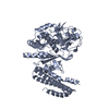
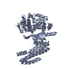
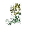
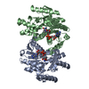
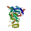
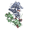
| Title | Structure of Thermus thermophilus CasA (Cse1) | ||||||
 Components Components | CSE1 | ||||||
 Keywords Keywords | IMMUNE SYSTEM / CRISPR / CASCADE / CASA | ||||||
| Function / homology | Topoisomerase I; Chain A, domain 4 - #100 / CRISPR-associated protein Cse1 / CRISPR-associated protein Cse1 (CRISPR_cse1) / Topoisomerase I; Chain A, domain 4 / defense response to virus / Orthogonal Bundle / Mainly Alpha / ACETATE ION / CRISPR-associated protein CasA/Cse1 Function and homology information Function and homology information | ||||||
| Biological species |   THERMUS THERMOPHILUS (bacteria) THERMUS THERMOPHILUS (bacteria) | ||||||
| Method |  X-RAY DIFFRACTION / SYNCHROTRON / MAD / Resolution: 2.3 Å X-RAY DIFFRACTION / SYNCHROTRON / MAD / Resolution: 2.3 Å | ||||||
 Authors Authors | Sashital, D.G. / Wiedenheft, B. / Doudna, J.A. | ||||||
 Citation Citation | Journal: Mol.Cell / Year: 2012 Title: Mechanism of Foreign DNA Selection in a Bacterial Adaptive Immune System. Authors: Sashital, D.G. / Wiedenheft, B. / Doudna, J.A. | ||||||
| History |
|
- Structure visualization
| Structure viewer | Molecule: MolmilJmol/JSmol |
|---|
- Downloads & links
-Download
| PDBx/mmCIF format | 4an8.cif.gz | 183.3 KB | Display | PDBx/mmCIF format |
|---|---|---|---|---|
| PDB format | pdb4an8.ent.gz | 147.6 KB | Display | PDB format |
| PDBx/mmJSON format | 4an8.json.gz | Tree view | PDBx/mmJSON format | |
| Others |  Other downloads Other downloads |
-Validation report
| Arichive directory | https://data.pdbj.org/pub/pdb/validation_reports/an/4an8ftp://data.pdbj.org/pub/pdb/validation_reports/an/4an8 | HTTPS FTP |
|---|
-Related structure data
| Similar structure data |
|---|


-Links
 PDBj
PDBj
- Assembly
| Deposited unit | 
| ||||||||
|---|---|---|---|---|---|---|---|---|---|
| 1 | 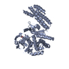
| ||||||||
| 2 | 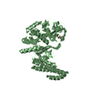
| ||||||||
| Unit cell |
|
-Components
| #1: Protein | Mass: 56545.379 Da / Num. of mol.: 2 Source method: isolated from a genetically manipulated source Details: TEV PROTEASE SCAR AT N-TERMINUS (GAGS) / Source: (gene. exp.) THERMUS THERMOPHILUS (bacteria) / Strain: HB8 / Gene: TTHB188 / Plasmid: PSV272 (PET VECTOR WITH ADDED TEV PROTEASE SITE) / Production host: #2: Chemical |   Mass: 59.044 Da / Num. of mol.: 3 / Source method: obtained synthetically / Formula: C2H3O2 Mass: 59.044 Da / Num. of mol.: 3 / Source method: obtained synthetically / Formula: C2H3O2#3: Water | ChemComp-HOH / |  Mass: 18.015 Da / Num. of mol.: 348 / Source method: isolated from a natural source / Formula: H2O Mass: 18.015 Da / Num. of mol.: 348 / Source method: isolated from a natural source / Formula: H2OHas protein modification | Y | |
|---|
-Experimental details
-Experiment
| Experiment | Method: X-RAY DIFFRACTION / Number of used crystals: 1 |
|---|
- Sample preparation
| Crystal | Density Matthews: 2.6 Å3/Da / Density % sol: 53 % / Description: NONE |
|---|---|
| Crystal grow | pH: 6.5 / Details: 2.1M NAAC, MES PH 6.5 |
-Data collection
| Diffraction | Mean temperature: 100 K |
|---|---|
| Diffraction source | Source: SYNCHROTRON / Site: ALS  / Beamline: 8.3.1 / Wavelength: 1.11587 / Beamline: 8.3.1 / Wavelength: 1.11587 |
| Detector | Type: ADSC CCD / Detector: CCD / Date: Mar 27, 2011 |
| Radiation | Protocol: SINGLE WAVELENGTH / Monochromatic (M) / Laue (L): M / Scattering type: x-ray |
| Radiation wavelength | Wavelength: 1.11587 Å / Relative weight: 1 |
| Reflection | Resolution: 2.3→46.83 Å / Num. obs: 51896 / % possible obs: 99.6 % / Observed criterion σ(I): 2.5 / Redundancy: 3.9 % / Biso Wilson estimate: 36.16 Å2 / Rmerge(I) obs: 0.06 / Net I/σ(I): 18.9 |
| Reflection shell | Resolution: 2.3→2.36 Å / Redundancy: 3.6 % / Rmerge(I) obs: 0.46 / Mean I/σ(I) obs: 2.9 / % possible all: 96.8 |
- Processing
| Software |
| ||||||||||||||||||||||||||||||||||||||||||||||||||||||||||||||||||||||||||||||||||||||||||||||||||||||||||||||||||||||||||||||||||||||||||||
|---|---|---|---|---|---|---|---|---|---|---|---|---|---|---|---|---|---|---|---|---|---|---|---|---|---|---|---|---|---|---|---|---|---|---|---|---|---|---|---|---|---|---|---|---|---|---|---|---|---|---|---|---|---|---|---|---|---|---|---|---|---|---|---|---|---|---|---|---|---|---|---|---|---|---|---|---|---|---|---|---|---|---|---|---|---|---|---|---|---|---|---|---|---|---|---|---|---|---|---|---|---|---|---|---|---|---|---|---|---|---|---|---|---|---|---|---|---|---|---|---|---|---|---|---|---|---|---|---|---|---|---|---|---|---|---|---|---|---|---|---|---|
| Refinement | Method to determine structure: MAD Starting model: NONE Resolution: 2.3→46.825 Å / SU ML: 0.67 / σ(F): 1.99 / Phase error: 23.33 / Stereochemistry target values: ML Details: CHAIN A RESIDUES 1-3, 130-143, 401-411 AND 497-502 ARE DISORDERED. CHAIN B RESIDUES 1-4, 129-143, 401-423 AND 494-502 ARE DISORDERED
| ||||||||||||||||||||||||||||||||||||||||||||||||||||||||||||||||||||||||||||||||||||||||||||||||||||||||||||||||||||||||||||||||||||||||||||
| Solvent computation | Shrinkage radii: 1.06 Å / VDW probe radii: 1.3 Å / Solvent model: FLAT BULK SOLVENT MODEL / Bsol: 48.448 Å2 / ksol: 0.372 e/Å3 | ||||||||||||||||||||||||||||||||||||||||||||||||||||||||||||||||||||||||||||||||||||||||||||||||||||||||||||||||||||||||||||||||||||||||||||
| Displacement parameters | Biso mean: 44.4 Å2
| ||||||||||||||||||||||||||||||||||||||||||||||||||||||||||||||||||||||||||||||||||||||||||||||||||||||||||||||||||||||||||||||||||||||||||||
| Refinement step | Cycle: LAST / Resolution: 2.3→46.825 Å
| ||||||||||||||||||||||||||||||||||||||||||||||||||||||||||||||||||||||||||||||||||||||||||||||||||||||||||||||||||||||||||||||||||||||||||||
| Refine LS restraints |
| ||||||||||||||||||||||||||||||||||||||||||||||||||||||||||||||||||||||||||||||||||||||||||||||||||||||||||||||||||||||||||||||||||||||||||||
| LS refinement shell |
|
