 Movie
Movie Controller

[English] 日本語
 Yorodumi
- PDB-4alb: Structure of Phenolic Acid Decarboxylase from Bacillus subtilis: ... -
+ Open data

- Basic information
| Entry | Database: PDB / ID: 4alb | ||||||
|---|---|---|---|---|---|---|---|
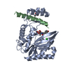
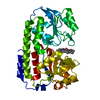
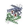
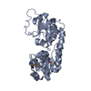
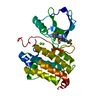
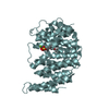
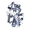
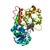
| Title | Structure of Phenolic Acid Decarboxylase from Bacillus subtilis: Tyr19Ala mutant in complex with coumaric acid | ||||||
 Components Components | PHENOLIC ACID DECARBOXYLASE PADC | ||||||
 Keywords Keywords | LYASE / LIPOCALIN OLD / CINNAMATE | ||||||
| Function / homology |  Function and homology information Function and homology informationphenacrylate decarboxylase / carboxy-lyase activity / response to toxic substance Similarity search - Function | ||||||
| Biological species |  | ||||||
| Method |  X-RAY DIFFRACTION / SYNCHROTRON / MOLECULAR REPLACEMENT / Resolution: 3.03 Å X-RAY DIFFRACTION / SYNCHROTRON / MOLECULAR REPLACEMENT / Resolution: 3.03 Å | ||||||
 Authors Authors | Frank, A. / Eborall, W. / Hyde, R. / Hart, S. / Turkenburg, J.P. / Grogan, G. | ||||||
 Citation Citation | Journal: Catal.Sci.Technol. / Year: 2012 Title: Mutational Analysis of Phenolic Acid Decarboxylase from Bacillus Subtilis (Bspad), which Converts Bio-Derived Phenolic Acids to Styrene Derivatives Authors: Frank, A. / Eborall, W. / Hyde, R. / Hart, S. / Turkenburg, J.P. / Grogan, G. | ||||||
| History |
|
- Structure visualization
| Structure viewer | Molecule: MolmilJmol/JSmol |
|---|
- Downloads & links
-Download
| PDBx/mmCIF format | 4alb.cif.gz | 107.7 KB | Display | PDBx/mmCIF format |
|---|---|---|---|---|
| PDB format | pdb4alb.ent.gz | 82.9 KB | Display | PDB format |
| PDBx/mmJSON format | 4alb.json.gz | Tree view | PDBx/mmJSON format | |
| Others |  Other downloads Other downloads |
-Validation report
| Arichive directory | https://data.pdbj.org/pub/pdb/validation_reports/al/4albftp://data.pdbj.org/pub/pdb/validation_reports/al/4alb | HTTPS FTP |
|---|
-Related structure data
| Related structure data |  2p8gS S: Starting model for refinement |
|---|---|
| Similar structure data |


-Links
 PDBj
PDBj


- Assembly
| Deposited unit | 
| ||||||||||||
|---|---|---|---|---|---|---|---|---|---|---|---|---|---|
| 1 | 
| ||||||||||||
| 2 | 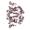
| ||||||||||||
| Unit cell |
| ||||||||||||
| Noncrystallographic symmetry (NCS) | NCS oper:
|
-Components
| #1: Protein | Mass: 19008.393 Da / Num. of mol.: 3 / Mutation: YES Source method: isolated from a genetically manipulated source Source: (gene. exp.) References: UniProt: O07006, Lyases; Carbon-carbon lyases; Carboxy-lyases #2: Chemical |   Mass: 164.158 Da / Num. of mol.: 3 / Source method: obtained synthetically / Formula: C9H8O3 Mass: 164.158 Da / Num. of mol.: 3 / Source method: obtained synthetically / Formula: C9H8O3#3: Water | ChemComp-HOH / |  Mass: 18.015 Da / Num. of mol.: 13 / Source method: isolated from a natural source / Formula: H2O Mass: 18.015 Da / Num. of mol.: 13 / Source method: isolated from a natural source / Formula: H2O |
|---|
-Experimental details
-Experiment
| Experiment | Method: X-RAY DIFFRACTION / Number of used crystals: 1 |
|---|
- Sample preparation
| Crystal | Density Matthews: 2.8 Å3/Da / Density % sol: 55 % / Description: NONE |
|---|---|
| Crystal grow | pH: 9 Details: 0.2 M KSCN, 17% (W/V) PEG 1000, 17% (W/V) PEG 8000, IN 0.1 M TRIS/HCL BUFFER AT PH 9. 15 MM COUMARIC ACID. PROTEIN AT 20 MG PER ML |
-Data collection
| Diffraction | Mean temperature: 120 K |
|---|---|
| Diffraction source | Source: SYNCHROTRON / Site: Diamond  / Beamline: I04 / Wavelength: 0.9763 / Beamline: I04 / Wavelength: 0.9763 |
| Detector | Type: ADSC CCD / Detector: CCD / Date: Oct 23, 2010 |
| Radiation | Protocol: SINGLE WAVELENGTH / Monochromatic (M) / Laue (L): M / Scattering type: x-ray |
| Radiation wavelength | Wavelength: 0.9763 Å / Relative weight: 1 |
| Reflection | Resolution: 3.02→53.6 Å / Num. obs: 12362 / % possible obs: 99.5 % / Observed criterion σ(I): 2 / Redundancy: 10.8 % / Rmerge(I) obs: 0.11 / Net I/σ(I): 21.4 |
| Reflection shell | Resolution: 3.02→3.1 Å / Redundancy: 11 % / Rmerge(I) obs: 0.86 / Mean I/σ(I) obs: 3.3 / % possible all: 98.6 |
- Processing
| Software |
| ||||||||||||||||||||||||||||||||||||||||||||||||||||||||||||||||||||||||||||||||||||||||||||||||||||||||||||||||||||||||||||||||||||||||||||||||||||||||||||||||||||||||||||||||||||||
|---|---|---|---|---|---|---|---|---|---|---|---|---|---|---|---|---|---|---|---|---|---|---|---|---|---|---|---|---|---|---|---|---|---|---|---|---|---|---|---|---|---|---|---|---|---|---|---|---|---|---|---|---|---|---|---|---|---|---|---|---|---|---|---|---|---|---|---|---|---|---|---|---|---|---|---|---|---|---|---|---|---|---|---|---|---|---|---|---|---|---|---|---|---|---|---|---|---|---|---|---|---|---|---|---|---|---|---|---|---|---|---|---|---|---|---|---|---|---|---|---|---|---|---|---|---|---|---|---|---|---|---|---|---|---|---|---|---|---|---|---|---|---|---|---|---|---|---|---|---|---|---|---|---|---|---|---|---|---|---|---|---|---|---|---|---|---|---|---|---|---|---|---|---|---|---|---|---|---|---|---|---|---|---|
| Refinement | Method to determine structure: MOLECULAR REPLACEMENT Starting model: PDB MODEL 2P8G Resolution: 3.03→92.85 Å / Cor.coef. Fo:Fc: 0.931 / Cor.coef. Fo:Fc free: 0.9 / SU B: 17.885 / SU ML: 0.317 / Cross valid method: THROUGHOUT / ESU R Free: 0.469 / Stereochemistry target values: MAXIMUM LIKELIHOOD / Details: HYDROGENS HAVE BEEN ADDED IN THE RIDING POSITIONS.
| ||||||||||||||||||||||||||||||||||||||||||||||||||||||||||||||||||||||||||||||||||||||||||||||||||||||||||||||||||||||||||||||||||||||||||||||||||||||||||||||||||||||||||||||||||||||
| Solvent computation | Ion probe radii: 0.8 Å / Shrinkage radii: 0.8 Å / VDW probe radii: 1.2 Å / Solvent model: MASK | ||||||||||||||||||||||||||||||||||||||||||||||||||||||||||||||||||||||||||||||||||||||||||||||||||||||||||||||||||||||||||||||||||||||||||||||||||||||||||||||||||||||||||||||||||||||
| Displacement parameters | Biso mean: 44.643 Å2
| ||||||||||||||||||||||||||||||||||||||||||||||||||||||||||||||||||||||||||||||||||||||||||||||||||||||||||||||||||||||||||||||||||||||||||||||||||||||||||||||||||||||||||||||||||||||
| Refinement step | Cycle: LAST / Resolution: 3.03→92.85 Å
| ||||||||||||||||||||||||||||||||||||||||||||||||||||||||||||||||||||||||||||||||||||||||||||||||||||||||||||||||||||||||||||||||||||||||||||||||||||||||||||||||||||||||||||||||||||||
| Refine LS restraints |
|