| Entry | Database: PDB / ID: 4aks
|
|---|
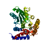
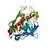
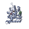
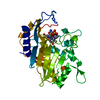
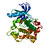
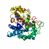
| Title | PatG macrocyclase domain |
|---|
 Components Components | THIAZOLINE OXIDASE/SUBTILISIN-LIKE PROTEASE |
|---|
 Keywords Keywords | HYDROLASE / PATELLAMIDE |
|---|
| Function / homology |  Function and homology information Function and homology information
ThcOx helix turn helix domain / : / ThcOx helix turn helix domain / Cyanobactin oxidase ThcOx, / Peptidase S8A, PatG / PatG/PatA-like domain / PatG domain / PatG, C-terminal / PatG Domain / PatG C-terminal ...ThcOx helix turn helix domain / : / ThcOx helix turn helix domain / Cyanobactin oxidase ThcOx, / Peptidase S8A, PatG / PatG/PatA-like domain / PatG domain / PatG, C-terminal / PatG Domain / PatG C-terminal / SagB-type dehydrogenase domain / : / Nitroreductase / Nitroreductase family / Nitroreductase-like / Peptidase S8/S53 domain / Serine proteases, subtilase family, serine active site. / Peptidase S8, subtilisin, Ser-active site / Serine proteases, subtilase domain profile. / Peptidase S8/S53 domain superfamily / Subtilase family / Peptidase S8/S53 domain / Rossmann fold / 3-Layer(aba) Sandwich / Alpha BetaSimilarity search - Domain/homology |
|---|
| Biological species |  PROCHLORON DIDEMNI (bacteria) PROCHLORON DIDEMNI (bacteria) |
|---|
| Method |  X-RAY DIFFRACTION / MOLECULAR REPLACEMENT / Resolution: 2.19 Å X-RAY DIFFRACTION / MOLECULAR REPLACEMENT / Resolution: 2.19 Å |
|---|
 Authors Authors | Koehnke, J. / Bent, A. / Houssen, W.E. / Zollman, D. / Morawitz, F. / Shirran, S. / Vendome, J. / Nneoyiegbe, A.F. / Trembleau, L. / Botting, C.H. ...Koehnke, J. / Bent, A. / Houssen, W.E. / Zollman, D. / Morawitz, F. / Shirran, S. / Vendome, J. / Nneoyiegbe, A.F. / Trembleau, L. / Botting, C.H. / Smith, M.C.M. / Jaspars, M. / Naismith, J.H. |
|---|
 Citation Citation | Journal: Nat.Struct.Mol.Biol. / Year: 2012
Title: The Mechanism of Patellamide Macrocyclization Revealed by the Characterization of the Patg Macrocyclase Domain.
Authors: Koehnke, J. / Bent, A. / Houssen, W.E. / Zollman, D. / Morawitz, F. / Shirran, S. / Vendome, J. / Nneoyiegbe, A.F. / Trembleau, L. / Botting, C.H. / Smith, M.C. / Jaspars, M. / Naismith, J.H. |
|---|
| History | | Deposition | Feb 28, 2012 | Deposition site: PDBE / Processing site: PDBE |
|---|
| Revision 1.0 | Jul 18, 2012 | Provider: repository / Type: Initial release |
|---|
| Revision 1.1 | Aug 22, 2012 | Group: Database references |
|---|
| Revision 1.2 | Nov 6, 2013 | Group: Structure summary |
|---|
| Revision 1.3 | Oct 23, 2024 | Group: Data collection / Database references ...Data collection / Database references / Other / Structure summary
Category: chem_comp_atom / chem_comp_bond ...chem_comp_atom / chem_comp_bond / database_2 / pdbx_database_status / pdbx_entry_details / pdbx_modification_feature
Item: _database_2.pdbx_DOI / _database_2.pdbx_database_accession / _pdbx_database_status.status_code_sf |
|---|
|
|---|
 Movie
Movie Controller


 Open data
 Basic information
 Structure visualization
 Downloads & links
 Other downloads
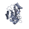


 PDBj
PDBj

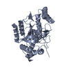
 Assembly

 Mass: 18.015 Da / Num. of mol.: 224 / Source method: isolated from a natural source / Formula: H2O
 Sample preparation
 Processing