 Movie
Movie Controller


+ Open data

- Basic information
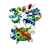
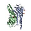
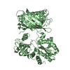
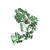
| Entry | Database: PDB / ID: 4ake | ||||||
|---|---|---|---|---|---|---|---|
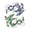
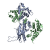
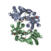
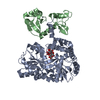
| Title | ADENYLATE KINASE | ||||||
 Components Components | ADENYLATE KINASE | ||||||
 Keywords Keywords | PHOSPHOTRANSFERASE / NUCLEOSIDE MONOPHOSPHATE KINASE / ATP:AMP PHOSPHOTRANSFERASE / MYOKINASE | ||||||
| Function / homology |  Function and homology information Function and homology informationpurine ribonucleotide interconversion / adenine metabolic process / nucleoside monophosphate metabolic process / ADP biosynthetic process / nucleoside diphosphate metabolic process / adenylate kinase / AMP kinase activity / AMP salvage / nucleoside diphosphate kinase activity / AMP binding ...purine ribonucleotide interconversion / adenine metabolic process / nucleoside monophosphate metabolic process / ADP biosynthetic process / nucleoside diphosphate metabolic process / adenylate kinase / AMP kinase activity / AMP salvage / nucleoside diphosphate kinase activity / AMP binding / magnesium ion binding / ATP binding / cytoplasm / cytosol Similarity search - Function | ||||||
| Biological species |  | ||||||
| Method |  X-RAY DIFFRACTION / Resolution: 2.2 Å X-RAY DIFFRACTION / Resolution: 2.2 Å | ||||||
 Authors Authors | Schlauderer, G.J. / Schulz, G.E. | ||||||
 Citation Citation | Journal: Structure / Year: 1996 Title: Adenylate kinase motions during catalysis: an energetic counterweight balancing substrate binding. Authors: Muller, C.W. / Schlauderer, G.J. / Reinstein, J. / Schulz, G.E. #1: Journal: Structure / Year: 1995Title: Movie of the Structural Changes During a Catalytic Cycle of Nucleoside Monophosphate Kinases Authors: Vonrhein, C. / Schlauderer, G.J. / Schulz, G.E. #2: Journal: J.Mol.Biol. / Year: 1992Title: Structure of the Complex between Adenylate Kinase from Escherichia Coli and the Inhibitor Ap5A Refined at 1.9 A Resolution. A Model for a Catalytic Transition State Authors: Muller, C.W. / Schulz, G.E. | ||||||
| History |
|
- Structure visualization
| Structure viewer | Molecule: MolmilJmol/JSmol |
|---|
- Downloads & links
-Download
| PDBx/mmCIF format | 4ake.cif.gz | 94.9 KB | Display | PDBx/mmCIF format |
|---|---|---|---|---|
| PDB format | pdb4ake.ent.gz | 73.3 KB | Display | PDB format |
| PDBx/mmJSON format | 4ake.json.gz | Tree view | PDBx/mmJSON format | |
| Others |  Other downloads Other downloads |
-Validation report
| Summary document | 4ake_validation.pdf.gz | 419.9 KB | Display | wwPDB validaton report |
|---|---|---|---|---|
| Full document | 4ake_full_validation.pdf.gz | 429 KB | Display | |
| Data in XML | 4ake_validation.xml.gz | 19.1 KB | Display | |
| Data in CIF | 4ake_validation.cif.gz | 26.7 KB | Display | |
| Arichive directory | https://data.pdbj.org/pub/pdb/validation_reports/ak/4akeftp://data.pdbj.org/pub/pdb/validation_reports/ak/4ake | HTTPS FTP |
-Related structure data
| Similar structure data |
|---|


-Links
 PDBj
PDBj- Assembly
| Deposited unit | 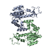
| ||||||||
|---|---|---|---|---|---|---|---|---|---|
| 1 |
| ||||||||
| Unit cell |
| ||||||||
| Noncrystallographic symmetry (NCS) | NCS oper: (Code: given Matrix: (0.999606, -0.012438, 0.025179), Vector: |
-Components
| #1: Protein | Mass: 23620.029 Da / Num. of mol.: 2 Source method: isolated from a genetically manipulated source Source: (gene. exp.) #2: Water | ChemComp-HOH / |  Mass: 18.015 Da / Num. of mol.: 147 / Source method: isolated from a natural source / Formula: H2O Mass: 18.015 Da / Num. of mol.: 147 / Source method: isolated from a natural source / Formula: H2OCompound details | THE SECONDARY STRUCTURAL ELEMENTS PRESENTED BELOW ARE FROM PROGRAM DSSP. FOR MANUAL ASSIGNMENTS ...THE SECONDARY STRUCTURAL | |
|---|
-Experimental details
-Experiment
| Experiment | Method: X-RAY DIFFRACTION |
|---|
- Sample preparation
| Crystal | Density Matthews: 2.36 Å3/Da / Density % sol: 48 % | |||||||||||||||||||||||||||||||||||||||||||||||||
|---|---|---|---|---|---|---|---|---|---|---|---|---|---|---|---|---|---|---|---|---|---|---|---|---|---|---|---|---|---|---|---|---|---|---|---|---|---|---|---|---|---|---|---|---|---|---|---|---|---|---|
| Crystal grow | *PLUS Temperature: 20 ℃ / Method: vapor diffusion, hanging drop | |||||||||||||||||||||||||||||||||||||||||||||||||
| Components of the solutions | *PLUS
|
-Data collection
| Diffraction source | Source: ROTATING ANODE / Wavelength: 1.5418 |
|---|---|
| Detector | Type: SIEMENS / Detector: AREA DETECTOR / Date: 1991 |
| Radiation | Monochromatic (M) / Laue (L): M / Scattering type: x-ray |
| Radiation wavelength | Wavelength: 1.5418 Å / Relative weight: 1 |
| Reflection | Resolution: 2.2→10 Å / Num. obs: 17932 / % possible obs: 82 % / Observed criterion σ(I): 0 / Redundancy: 3 % / Rmerge(I) obs: 0.063 |
| Reflection | *PLUS Rmerge(I) obs: 0.063 |
- Processing
| Software |
| ||||||||||||||||||||||||||||||||||||||||||||||||||||||||||||||||||||||||||||||||
|---|---|---|---|---|---|---|---|---|---|---|---|---|---|---|---|---|---|---|---|---|---|---|---|---|---|---|---|---|---|---|---|---|---|---|---|---|---|---|---|---|---|---|---|---|---|---|---|---|---|---|---|---|---|---|---|---|---|---|---|---|---|---|---|---|---|---|---|---|---|---|---|---|---|---|---|---|---|---|---|---|---|
| Refinement | Resolution: 2.2→10 Å / σ(F): 0
| ||||||||||||||||||||||||||||||||||||||||||||||||||||||||||||||||||||||||||||||||
| Displacement parameters | Biso mean: 42 Å2 | ||||||||||||||||||||||||||||||||||||||||||||||||||||||||||||||||||||||||||||||||
| Refine analyze | Luzzati coordinate error obs: 0.3 Å | ||||||||||||||||||||||||||||||||||||||||||||||||||||||||||||||||||||||||||||||||
| Refinement step | Cycle: LAST / Resolution: 2.2→10 Å
| ||||||||||||||||||||||||||||||||||||||||||||||||||||||||||||||||||||||||||||||||
| Refine LS restraints |
| ||||||||||||||||||||||||||||||||||||||||||||||||||||||||||||||||||||||||||||||||
| Refinement | *PLUS Rfactor obs: 0.183 / Rfactor Rwork: 0.183 | ||||||||||||||||||||||||||||||||||||||||||||||||||||||||||||||||||||||||||||||||
| Solvent computation | *PLUS | ||||||||||||||||||||||||||||||||||||||||||||||||||||||||||||||||||||||||||||||||
| Displacement parameters | *PLUS |
