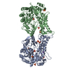
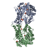
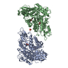
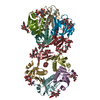
A: C-TYPE LECTIN DOMAIN FAMILY 4 MEMBER K B: C-TYPE LECTIN DOMAIN FAMILY 4 MEMBER K C: C-TYPE LECTIN DOMAIN FAMILY 4 MEMBER K D: C-TYPE LECTIN DOMAIN FAMILY 4 MEMBER K hetero molecules
A: C-TYPE LECTIN DOMAIN FAMILY 4 MEMBER K B: C-TYPE LECTIN DOMAIN FAMILY 4 MEMBER K C: C-TYPE LECTIN DOMAIN FAMILY 4 MEMBER K D: C-TYPE LECTIN DOMAIN FAMILY 4 MEMBER K hetero molecules
A: C-TYPE LECTIN DOMAIN FAMILY 4 MEMBER K B: C-TYPE LECTIN DOMAIN FAMILY 4 MEMBER K C: C-TYPE LECTIN DOMAIN FAMILY 4 MEMBER K D: C-TYPE LECTIN DOMAIN FAMILY 4 MEMBER K hetero molecules
Mass: 18.015 Da / Num. of mol.: 807 / Source method: isolated from a natural source / Formula: H2O
Compound details
ENGINEERED RESIDUE IN CHAIN A, PHE 241 TO LEU ENGINEERED RESIDUE IN CHAIN B, PHE 241 TO LEU ...ENGINEERED RESIDUE IN CHAIN A, PHE 241 TO LEU ENGINEERED RESIDUE IN CHAIN B, PHE 241 TO LEU ENGINEERED RESIDUE IN CHAIN C, PHE 241 TO LEU ENGINEERED RESIDUE IN CHAIN D, PHE 241 TO LEU
Has protein modification
Y
-
Experimental details
-
Experiment
Experiment
Method: X-RAY DIFFRACTION / Number of used crystals: 1
-
Sample preparation
Crystal
Density Matthews: 2.02 Å3/Da / Density % sol: 39.2 % / Description: NONE
Crystal grow
Method: vapor diffusion, hanging drop / pH: 6 Details: HANGING DROP METHOD 100 MM MES PH 6.0, 200 MM MGCL2 AND 17% PEG 3350
 Movie
Movie Controller

 Yorodumi
 Open data
 Basic information
 Components
 Keywords
 Function and homology information
 HOMO SAPIENS (human)
 X-RAY DIFFRACTION /
  Authors
 Citation
 Structure visualization
 Downloads & links
 Other downloads


 PDBj
PDBj


 Assembly


 Mass: 40.078 Da / Num. of mol.: 4 / Source method: obtained synthetically / Formula: Ca
 Mass: 24.305 Da / Num. of mol.: 4 / Source method: obtained synthetically / Formula: Mg
 Mass: 35.453 Da / Num. of mol.: 1 / Source method: obtained synthetically / Formula: Cl
 Mass: 18.015 Da / Num. of mol.: 807 / Source method: isolated from a natural source / Formula: H2O
 Sample preparation
 / Beamline: ID14-2 / Wavelength: 0.933
  Processing