 Movie
Movie Controller

[English] 日本語
 Yorodumi
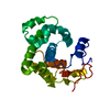
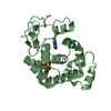
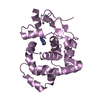
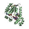
- PDB-4aia: The structural basis of 3-methyladenine recognition by 3- methyla... -
+ Open data

- Basic information
| Entry | Database: PDB / ID: 4aia | ||||||
|---|---|---|---|---|---|---|---|
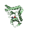
| Title | The structural basis of 3-methyladenine recognition by 3- methyladenine DNA glycosylase I (TAG) from Staphylococcus aureus | ||||||
 Components Components | DNA-3-METHYLADENINE GLYCOSYLASE I | ||||||
 Keywords Keywords | HYDROLASE / 3-METHYLADENINE RECOGNITION / DNA REPAIR | ||||||
| Function / homology | Hypothetical protein; domain 2 / Endonuclease III; domain 1 / Orthogonal Bundle / Mainly Alpha / 3-METHYL-3H-PURIN-6-YLAMINE / :  Function and homology information Function and homology information | ||||||
| Biological species |   STAPHYLOCOCCUS AUREUS (bacteria) STAPHYLOCOCCUS AUREUS (bacteria) | ||||||
| Method |  X-RAY DIFFRACTION / SYNCHROTRON / MOLECULAR REPLACEMENT / Resolution: 1.8 Å X-RAY DIFFRACTION / SYNCHROTRON / MOLECULAR REPLACEMENT / Resolution: 1.8 Å | ||||||
 Authors Authors | Yan, X. / Naismith, J.H. | ||||||
 Citation Citation | Journal: Acta Crystallogr.,Sect.F / Year: 2012 Title: A Model for 3-Methyladenine Recognition by 3-Methyladenine DNA Glycosylase I (Tag) from Staphylococcus Aureus. Authors: Zhu, X. / Yan, X. / Carter, L.G. / Liu, H. / Graham, S. / Coote, P.J. / Naismith, J.H. | ||||||
| History |
|
- Structure visualization
| Structure viewer | Molecule: MolmilJmol/JSmol |
|---|
- Downloads & links
-Download
| PDBx/mmCIF format | 4aia.cif.gz | 400.8 KB | Display | PDBx/mmCIF format |
|---|---|---|---|---|
| PDB format | pdb4aia.ent.gz | 331.5 KB | Display | PDB format |
| PDBx/mmJSON format | 4aia.json.gz | Tree view | PDBx/mmJSON format | |
| Others |  Other downloads Other downloads |
-Validation report
| Summary document | 4aia_validation.pdf.gz | 483.4 KB | Display | wwPDB validaton report |
|---|---|---|---|---|
| Full document | 4aia_full_validation.pdf.gz | 488.8 KB | Display | |
| Data in XML | 4aia_validation.xml.gz | 44.7 KB | Display | |
| Data in CIF | 4aia_validation.cif.gz | 65.9 KB | Display | |
| Arichive directory | https://data.pdbj.org/pub/pdb/validation_reports/ai/4aiaftp://data.pdbj.org/pub/pdb/validation_reports/ai/4aia | HTTPS FTP |
-Related structure data
| Related structure data |  4ai4C  4ai5C  2jg6S S: Starting model for refinement C: citing same article ( |
|---|---|
| Similar structure data |


-Links
 PDBj
PDBj- Assembly
| Deposited unit | 
| |||||||||
|---|---|---|---|---|---|---|---|---|---|---|
| 1 | 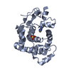
| |||||||||
| 2 | 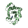
| |||||||||
| 3 | 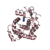
| |||||||||
| 4 | 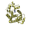
| |||||||||
| 5 | 
| |||||||||
| Unit cell |
| |||||||||
| Components on special symmetry positions |
|
-Components
| #1: Protein | Mass: 21573.484 Da / Num. of mol.: 5 Source method: isolated from a genetically manipulated source Source: (gene. exp.) STAPHYLOCOCCUS AUREUS (bacteria) / Strain: MSSA476 / Plasmid: PDEST14 / Production host: References: UniProt: Q6G8R1, DNA-3-methyladenine glycosylase I #2: Chemical | ChemComp-ZN /   Mass: 65.409 Da / Num. of mol.: 5 / Source method: obtained synthetically / Formula: Zn Mass: 65.409 Da / Num. of mol.: 5 / Source method: obtained synthetically / Formula: Zn#3: Chemical | ChemComp-ADK /   Mass: 149.153 Da / Num. of mol.: 5 / Source method: obtained synthetically / Formula: C6H7N5 Mass: 149.153 Da / Num. of mol.: 5 / Source method: obtained synthetically / Formula: C6H7N5#4: Chemical | ChemComp-SO4 /   Mass: 96.063 Da / Num. of mol.: 4 / Source method: obtained synthetically / Formula: SO4 Mass: 96.063 Da / Num. of mol.: 4 / Source method: obtained synthetically / Formula: SO4#5: Water | ChemComp-HOH / |  Mass: 18.015 Da / Num. of mol.: 927 / Source method: isolated from a natural source / Formula: H2O Mass: 18.015 Da / Num. of mol.: 927 / Source method: isolated from a natural source / Formula: H2O |
|---|
-Experimental details
-Experiment
| Experiment | Method: X-RAY DIFFRACTION / Number of used crystals: 1 |
|---|
- Sample preparation
| Crystal | Density Matthews: 2.46 Å3/Da / Density % sol: 50 % / Description: NONE |
|---|---|
| Crystal grow | pH: 8.5 / Details: pH 8.5 |
-Data collection
| Diffraction | Mean temperature: 100 K |
|---|---|
| Diffraction source | Source: SYNCHROTRON / Site: ESRF  / Beamline: ID14-2 / Wavelength: 0.933 / Beamline: ID14-2 / Wavelength: 0.933 |
| Detector | Type: QUANTUM 4 / Detector: CCD / Date: Sep 1, 2007 |
| Radiation | Protocol: SINGLE WAVELENGTH / Monochromatic (M) / Laue (L): M / Scattering type: x-ray |
| Radiation wavelength | Wavelength: 0.933 Å / Relative weight: 1 |
| Reflection | Resolution: 1.8→29.6 Å / Num. obs: 92544 / % possible obs: 98.4 % / Observed criterion σ(I): 2 / Redundancy: 3.7 % / Rmerge(I) obs: 0.05 / Net I/σ(I): 17.5 |
| Reflection shell | Resolution: 1.8→1.9 Å / Redundancy: 3.1 % / Rmerge(I) obs: 0.24 / Mean I/σ(I) obs: 4.8 / % possible all: 91.6 |
- Processing
| Software |
| ||||||||||||||||||||||||||||||||||||||||||||||||||||||||||||||||||||||||||||||||||||||||||||||||||||||||||||||||||||||||||||||||||||||||||||||||||||||||||||||||||||||||||||||||||||||
|---|---|---|---|---|---|---|---|---|---|---|---|---|---|---|---|---|---|---|---|---|---|---|---|---|---|---|---|---|---|---|---|---|---|---|---|---|---|---|---|---|---|---|---|---|---|---|---|---|---|---|---|---|---|---|---|---|---|---|---|---|---|---|---|---|---|---|---|---|---|---|---|---|---|---|---|---|---|---|---|---|---|---|---|---|---|---|---|---|---|---|---|---|---|---|---|---|---|---|---|---|---|---|---|---|---|---|---|---|---|---|---|---|---|---|---|---|---|---|---|---|---|---|---|---|---|---|---|---|---|---|---|---|---|---|---|---|---|---|---|---|---|---|---|---|---|---|---|---|---|---|---|---|---|---|---|---|---|---|---|---|---|---|---|---|---|---|---|---|---|---|---|---|---|---|---|---|---|---|---|---|---|---|---|
| Refinement | Method to determine structure: MOLECULAR REPLACEMENT Starting model: PDB ENTRY 2JG6 Resolution: 1.8→28.19 Å / Cor.coef. Fo:Fc: 0.947 / Cor.coef. Fo:Fc free: 0.929 / SU B: 4.544 / SU ML: 0.082 / Cross valid method: THROUGHOUT / ESU R: 0.133 / ESU R Free: 0.126 / Stereochemistry target values: MAXIMUM LIKELIHOOD Details: HYDROGENS HAVE BEEN ADDED IN THE RIDING POSITIONS.HYDROGENS HAVE BEEN USED IF PRESENT IN INPUT. U VALUES WITH TLS ADDED.
| ||||||||||||||||||||||||||||||||||||||||||||||||||||||||||||||||||||||||||||||||||||||||||||||||||||||||||||||||||||||||||||||||||||||||||||||||||||||||||||||||||||||||||||||||||||||
| Solvent computation | Ion probe radii: 0.8 Å / Shrinkage radii: 0.8 Å / VDW probe radii: 1.2 Å / Solvent model: MASK | ||||||||||||||||||||||||||||||||||||||||||||||||||||||||||||||||||||||||||||||||||||||||||||||||||||||||||||||||||||||||||||||||||||||||||||||||||||||||||||||||||||||||||||||||||||||
| Displacement parameters | Biso mean: 22.376 Å2
| ||||||||||||||||||||||||||||||||||||||||||||||||||||||||||||||||||||||||||||||||||||||||||||||||||||||||||||||||||||||||||||||||||||||||||||||||||||||||||||||||||||||||||||||||||||||
| Refinement step | Cycle: LAST / Resolution: 1.8→28.19 Å
| ||||||||||||||||||||||||||||||||||||||||||||||||||||||||||||||||||||||||||||||||||||||||||||||||||||||||||||||||||||||||||||||||||||||||||||||||||||||||||||||||||||||||||||||||||||||
| Refine LS restraints |
|