[English] 日本語
Yorodumi
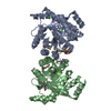
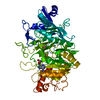
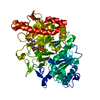
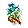
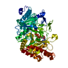
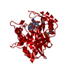
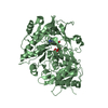
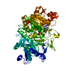
- PDB-4afy: Crystal structure of the FimX EAL domain in complex with reaction... -

+
Open data


ID or keywords:

Loading...

-
Basic information

Entry
Database: PDB / ID: 4afy
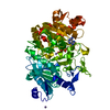
TitleCrystal structure of the FimX EAL domain in complex with reaction product pGpG
Components
  • 5'-R(*GP*GP)-3'
  • FIMX
KeywordsHYDROLASE / CDIGMP BIOFILM / PHOSPHODIESTERASE
Function / homology
Function and homology information


phosphorelay signal transduction system / nucleotide binding / regulation of DNA-templated transcription / identical protein binding / metal ion binding
Similarity search - Function
EAL domain / Putative diguanylate phosphodiesterase / EAL domain / EAL domain superfamily / EAL domain / EAL domain profile. / Diguanylate cyclase, GGDEF domain / diguanylate cyclase / GGDEF domain profile. / GGDEF domain ...EAL domain / Putative diguanylate phosphodiesterase / EAL domain / EAL domain superfamily / EAL domain / EAL domain profile. / Diguanylate cyclase, GGDEF domain / diguanylate cyclase / GGDEF domain profile. / GGDEF domain / Nucleotide cyclase / Signal transduction response regulator, receiver domain / Response regulatory domain profile. / CheY-like superfamily / PAS fold / PAS fold / PAS domain / PAS domain superfamily / Reverse transcriptase/Diguanylate cyclase domain / TIM Barrel / Alpha-Beta Barrel / Alpha Beta
Similarity search - Domain/homology
RNA / Uncharacterized protein / FimX
Similarity search - Component
Biological speciesPSEUDOMONAS AERUGINOSA PAO1 (bacteria)
SYNTHETIC CONSTRUCT (others)
MethodX-RAY DIFFRACTION / SYNCHROTRON / MOLECULAR REPLACEMENT / Resolution: 2.01 Å
AuthorsRobert-Paganin, J. / Nonin-Lecomte, S. / Rety, S.
CitationJournal: Plos One / Year: 2012
Title: Crystal Structure of an Eal Domain in Complex with Reaction Product 5'-Pgpg
Authors: Robert-Paganin, J. / Nonin-Lecomte, S. / Rety, S.
History
DepositionJan 23, 2012Deposition site: PDBE / Processing site: PDBE
Revision 1.0Jan 9, 2013Provider: repository / Type: Initial release
Revision 1.1Jan 30, 2013Group: Database references
Revision 1.2Oct 21, 2015Group: Source and taxonomy
Revision 1.3Dec 20, 2023Group: Data collection / Database references ...Data collection / Database references / Derived calculations / Other / Refinement description
Category: chem_comp_atom / chem_comp_bond ...chem_comp_atom / chem_comp_bond / database_2 / pdbx_database_status / pdbx_initial_refinement_model / pdbx_struct_conn_angle / struct_conn / struct_site
Item: _database_2.pdbx_DOI / _database_2.pdbx_database_accession ..._database_2.pdbx_DOI / _database_2.pdbx_database_accession / _pdbx_database_status.status_code_sf / _pdbx_struct_conn_angle.ptnr1_auth_asym_id / _pdbx_struct_conn_angle.ptnr1_auth_comp_id / _pdbx_struct_conn_angle.ptnr1_auth_seq_id / _pdbx_struct_conn_angle.ptnr1_label_asym_id / _pdbx_struct_conn_angle.ptnr1_label_atom_id / _pdbx_struct_conn_angle.ptnr1_label_comp_id / _pdbx_struct_conn_angle.ptnr1_label_seq_id / _pdbx_struct_conn_angle.ptnr2_auth_seq_id / _pdbx_struct_conn_angle.ptnr2_label_asym_id / _pdbx_struct_conn_angle.ptnr3_auth_asym_id / _pdbx_struct_conn_angle.ptnr3_auth_comp_id / _pdbx_struct_conn_angle.ptnr3_auth_seq_id / _pdbx_struct_conn_angle.ptnr3_label_asym_id / _pdbx_struct_conn_angle.ptnr3_label_atom_id / _pdbx_struct_conn_angle.ptnr3_label_comp_id / _pdbx_struct_conn_angle.ptnr3_label_seq_id / _pdbx_struct_conn_angle.value / _struct_conn.pdbx_dist_value / _struct_conn.ptnr1_auth_comp_id / _struct_conn.ptnr1_auth_seq_id / _struct_conn.ptnr1_label_asym_id / _struct_conn.ptnr1_label_atom_id / _struct_conn.ptnr1_label_comp_id / _struct_conn.ptnr1_label_seq_id / _struct_conn.ptnr2_auth_asym_id / _struct_conn.ptnr2_auth_comp_id / _struct_conn.ptnr2_auth_seq_id / _struct_conn.ptnr2_label_asym_id / _struct_conn.ptnr2_label_atom_id / _struct_conn.ptnr2_label_comp_id / _struct_conn.ptnr2_label_seq_id / _struct_site.pdbx_auth_asym_id / _struct_site.pdbx_auth_comp_id / _struct_site.pdbx_auth_seq_id
Remark 700 SHEET DETERMINATION METHOD: DSSP THE SHEETS PRESENTED AS "AA" IN EACH CHAIN ON SHEET RECORDS BELOW ... SHEET DETERMINATION METHOD: DSSP THE SHEETS PRESENTED AS "AA" IN EACH CHAIN ON SHEET RECORDS BELOW IS ACTUALLY AN 8-STRANDED BARREL THIS IS REPRESENTED BY A 9-STRANDED SHEET IN WHICH THE FIRST AND LAST STRANDS ARE IDENTICAL.

-
Structure visualization

Structure viewerMolecule:
MolmilJmol/JSmol

Downloads & links

-
Assembly

Deposited unit
A: FIMX
B: FIMX
C: 5'-R(*GP*GP)-3'
D: 5'-R(*GP*GP)-3'
hetero molecules


Theoretical massNumber of molelcules
Total (without water)61,9759
Polymers61,8564
Non-polymers1195
Water4,828268
1


  • Idetical with deposited unit
  • defined by author&software
TypeNameSymmetry operationNumber
identity operation1_555x,y,z1
Buried area5840 Å2
ΔGint-76.7 kcal/mol
Surface area21330 Å2
MethodPISA
Unit cell
Length a, b, c (Å)104.330, 104.330, 154.240
Angle α, β, γ (deg.)90.00, 90.00, 90.00
Int Tables number96
Space group name H-MP43212
Components on special symmetry positions
IDModelComponents
11A-2055-

HOH

21B-2037-

HOH

-
Components

#1: Protein FIMX


Mass: 30282.393 Da / Num. of mol.: 2 / Fragment: EAL DOMAIN, RESIDUES 439-691
Source method: isolated from a genetically manipulated source
Source: (gene. exp.) PSEUDOMONAS AERUGINOSA PAO1 (bacteria) / Gene: PA4959 / Production host: ESCHERICHIA COLI (E. coli) / Strain (production host): BL21 / Variant (production host): ROSETTA 2
References: UniProt: Q02F59, UniProt: Q9HUK6*PLUS, cyclic-guanylate-specific phosphodiesterase
#2: RNA chain 5'-R(*GP*GP)-3'


Mass: 645.454 Da / Num. of mol.: 2 / Source method: obtained synthetically / Source: (synth.) SYNTHETIC CONSTRUCT (others)
#3: Chemical ChemComp-MG / MAGNESIUM ION


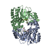
Mass: 24.305 Da / Num. of mol.: 3 / Source method: obtained synthetically / Formula: Mg
#4: Chemical ChemComp-NA / SODIUM ION


Mass: 22.990 Da / Num. of mol.: 2 / Source method: obtained synthetically / Formula: Na
#5: Water ChemComp-HOH / water


Mass: 18.015 Da / Num. of mol.: 268 / Source method: isolated from a natural source / Formula: H2O

-
Experimental details

-
Experiment

ExperimentMethod: X-RAY DIFFRACTION / Number of used crystals: 1

-
Sample preparation

CrystalDensity Matthews: 3.47 Å3/Da / Density % sol: 64.6 % / Description: NONE
Crystal growpH: 7 / Details: HEPES 0.1 M PH 8.0, NAH2PO4 0.8 M K2HPO4 0.8 M

-
Data collection

DiffractionMean temperature: 100 K
Diffraction sourceSource: SYNCHROTRON / Site: SOLEIL / Beamline: PROXIMA 1 / Wavelength: 0.9793
DetectorType: ADSC QUANTUM 315r / Detector: CCD / Date: Dec 4, 2008
RadiationProtocol: SINGLE WAVELENGTH / Monochromatic (M) / Laue (L): M / Scattering type: x-ray
Radiation wavelengthWavelength: 0.9793 Å / Relative weight: 1
ReflectionResolution: 2.01→50 Å / Num. obs: 57232 / % possible obs: 99.9 % / Observed criterion σ(I): 2 / Redundancy: 13.6 % / Biso Wilson estimate: 43.8 Å2 / Rmerge(I) obs: 0.06 / Net I/σ(I): 30.6
Reflection shellResolution: 2.01→2.06 Å / Redundancy: 8.7 % / Rmerge(I) obs: 0.38 / Mean I/σ(I) obs: 5.8 / % possible all: 99.5

-
Processing

Software
NameVersionClassification
PHENIX(PHENIX.REFINE)refinement
XDSdata reduction
XDSdata scaling
BALBESphasing
RefinementMethod to determine structure: MOLECULAR REPLACEMENT
Starting model: PDB ENTRY 2R6O
Resolution: 2.01→43.209 Å / SU ML: 0.29 / σ(F): 1.99 / Phase error: 23.27 / Stereochemistry target values: ML
RfactorNum. reflection% reflection
Rfree0.239 2890 5.1 %
Rwork0.2085 --
obs0.2101 57230 99.95 %
Solvent computationShrinkage radii: 0.98 Å / VDW probe radii: 1.2 Å / Solvent model: FLAT BULK SOLVENT MODEL / Bsol: 44.985 Å2 / ksol: 0.357 e/Å3
Displacement parametersBiso mean: 46.8 Å2
Baniso -1Baniso -2Baniso -3
1-0.5263 Å20 Å20 Å2
2--0.5263 Å20 Å2
3----1.0526 Å2
Refinement stepCycle: LAST / Resolution: 2.01→43.209 Å
ProteinNucleic acidLigandSolventTotal
Num. atoms3912 92 5 268 4277
Refine LS restraints
Refine-IDTypeDev idealNumber
X-RAY DIFFRACTIONf_bond_d0.0144097
X-RAY DIFFRACTIONf_angle_d1.4075581
X-RAY DIFFRACTIONf_dihedral_angle_d15.9521503
X-RAY DIFFRACTIONf_chiral_restr0.077639
X-RAY DIFFRACTIONf_plane_restr0.007708
LS refinement shell
Resolution (Å)Rfactor RfreeNum. reflection RfreeRfactor RworkNum. reflection RworkRefine-ID% reflection obs (%)
2.0101-2.0430.29051410.27942510X-RAY DIFFRACTION99
2.043-2.07830.29711260.24782554X-RAY DIFFRACTION100
2.0783-2.11610.30361180.24862573X-RAY DIFFRACTION100
2.1161-2.15680.28061230.24212537X-RAY DIFFRACTION100
2.1568-2.20080.32251360.24332566X-RAY DIFFRACTION100
2.2008-2.24860.28691360.23892549X-RAY DIFFRACTION100
2.2486-2.30090.28051190.22462561X-RAY DIFFRACTION100
2.3009-2.35850.31321140.21682589X-RAY DIFFRACTION100
2.3585-2.42220.26281320.23512572X-RAY DIFFRACTION100
2.4222-2.49350.27741490.22032556X-RAY DIFFRACTION100
2.4935-2.5740.27251450.2222531X-RAY DIFFRACTION100
2.574-2.6660.26121500.21152575X-RAY DIFFRACTION100
2.666-2.77270.24111460.21692566X-RAY DIFFRACTION100
2.7727-2.89890.23431350.20982584X-RAY DIFFRACTION100
2.8989-3.05170.25171340.21912599X-RAY DIFFRACTION100
3.0517-3.24280.22251470.20872573X-RAY DIFFRACTION100
3.2428-3.49310.24231660.20912583X-RAY DIFFRACTION100
3.4931-3.84440.24471330.20382620X-RAY DIFFRACTION100
3.8444-4.40020.19061510.17342636X-RAY DIFFRACTION100
4.4002-5.5420.20531490.17422669X-RAY DIFFRACTION100
5.542-43.21850.2331400.22172837X-RAY DIFFRACTION100

+
About Yorodumi

-
News

-
Feb 9, 2022. New format data for meta-information of EMDB entries

New format data for meta-information of EMDB entries

  • Version 3 of the EMDB header file is now the official format.
  • The previous official version 1.9 will be removed from the archive.

Related info.:EMDB header

External links:wwPDB to switch to version 3 of the EMDB data model

-
Aug 12, 2020. Covid-19 info

Covid-19 info

URL: https://pdbjlvh1.pdbj.org/emnavi/covid19.php

New page: Covid-19 featured information page in EM Navigator.

Related info.:Covid-19 info / Mar 5, 2020. Novel coronavirus structure data

+
Mar 5, 2020. Novel coronavirus structure data

Novel coronavirus structure data

Related info.:Yorodumi Speices / Aug 12, 2020. Covid-19 info

External links:COVID-19 featured content - PDBj / Molecule of the Month (242):Coronavirus Proteases

+
Jan 31, 2019. EMDB accession codes are about to change! (news from PDBe EMDB page)

EMDB accession codes are about to change! (news from PDBe EMDB page)

  • The allocation of 4 digits for EMDB accession codes will soon come to an end. Whilst these codes will remain in use, new EMDB accession codes will include an additional digit and will expand incrementally as the available range of codes is exhausted. The current 4-digit format prefixed with “EMD-” (i.e. EMD-XXXX) will advance to a 5-digit format (i.e. EMD-XXXXX), and so on. It is currently estimated that the 4-digit codes will be depleted around Spring 2019, at which point the 5-digit format will come into force.
  • The EM Navigator/Yorodumi systems omit the EMD- prefix.

Related info.:Q: What is EMD? / ID/Accession-code notation in Yorodumi/EM Navigator

External links:EMDB Accession Codes are Changing Soon! / Contact to PDBj

+
Jul 12, 2017. Major update of PDB

Major update of PDB

  • wwPDB released updated PDB data conforming to the new PDBx/mmCIF dictionary.
  • This is a major update changing the version number from 4 to 5, and with Remediation, in which all the entries are updated.
  • In this update, many items about electron microscopy experimental information are reorganized (e.g. em_software).
  • Now, EM Navigator and Yorodumi are based on the updated data.

External links:wwPDB Remediation / Enriched Model Files Conforming to OneDep Data Standards Now Available in the PDB FTP Archive

-
Yorodumi

Thousand views of thousand structures

  • Yorodumi is a browser for structure data from EMDB, PDB, SASBDB, etc.
  • This page is also the successor to EM Navigator detail page, and also detail information page/front-end page for Omokage search.
  • The word "yorodu" (or yorozu) is an old Japanese word meaning "ten thousand". "mi" (miru) is to see.

Related info.:EMDB / PDB / SASBDB / Comparison of 3 databanks / Yorodumi Search / Aug 31, 2016. New EM Navigator & Yorodumi / Yorodumi Papers / Jmol/JSmol / Function and homology information / Changes in new EM Navigator and Yorodumi

Read more