 Movie
Movie Controller

[English] 日本語
 Yorodumi
- PDB-4ade: Structural and functional study of succinyl-ornithine transaminas... -
+ Open data

- Basic information
| Entry | Database: PDB / ID: 4ade | ||||||
|---|---|---|---|---|---|---|---|
| Title | Structural and functional study of succinyl-ornithine transaminase from E. coli | ||||||
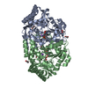
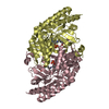
 Components Components | SUCCINYLORNITHINE TRANSAMINASE | ||||||
 Keywords Keywords | TRANSFERASE / PLP ENZYMES / AMINOTRANSFERASE | ||||||
| Function / homology |  Function and homology information Function and homology informationsuccinylornithine transaminase / succinylornithine transaminase activity / L-ornithine catabolic process / L-arginine catabolic process to succinate / N2-acetyl-L-ornithine:2-oxoglutarate 5-aminotransferase activity / L-arginine catabolic process to L-glutamate / L-arginine biosynthetic process via ornithine / L-arginine catabolic process / pyridoxal phosphate binding / identical protein binding Similarity search - Function | ||||||
| Biological species |  | ||||||
| Method |  X-RAY DIFFRACTION / SYNCHROTRON / MOLECULAR REPLACEMENT / Resolution: 2.75 Å X-RAY DIFFRACTION / SYNCHROTRON / MOLECULAR REPLACEMENT / Resolution: 2.75 Å | ||||||
 Authors Authors | Newman, J. / Peat, T.S. | ||||||
 Citation Citation | Journal: Plos One / Year: 2013 Title: Determination of the Structure of the Catabolic N-Succinylornithine Transaminase (Astc) from Escherichia Coli. Authors: Newman, J. / Seabrook, S. / Surjadi, R. / Williams, C.C. / Lucent, D. / Wilding, M. / Scott, C. / Peat, T.S. | ||||||
| History |
|
- Structure visualization
| Structure viewer | Molecule: MolmilJmol/JSmol |
|---|
- Downloads & links
-Download
| PDBx/mmCIF format | 4ade.cif.gz | 151.2 KB | Display | PDBx/mmCIF format |
|---|---|---|---|---|
| PDB format | pdb4ade.ent.gz | 120.3 KB | Display | PDB format |
| PDBx/mmJSON format | 4ade.json.gz | Tree view | PDBx/mmJSON format | |
| Others |  Other downloads Other downloads |
-Validation report
| Summary document | 4ade_validation.pdf.gz | 433.9 KB | Display | wwPDB validaton report |
|---|---|---|---|---|
| Full document | 4ade_full_validation.pdf.gz | 445.5 KB | Display | |
| Data in XML | 4ade_validation.xml.gz | 28.1 KB | Display | |
| Data in CIF | 4ade_validation.cif.gz | 38.6 KB | Display | |
| Arichive directory | https://data.pdbj.org/pub/pdb/validation_reports/ad/4adeftp://data.pdbj.org/pub/pdb/validation_reports/ad/4ade | HTTPS FTP |
-Related structure data
| Related structure data | 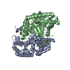 4adbC  4adcC 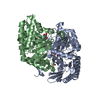 4addC  2pb2S C: citing same article ( S: Starting model for refinement |
|---|---|
| Similar structure data |


-Links
 PDBj
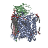
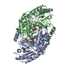
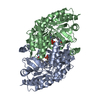
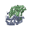
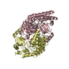
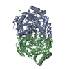
PDBj- Assembly
| Deposited unit | 
| |||||||||
|---|---|---|---|---|---|---|---|---|---|---|
| 1 | 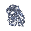
| |||||||||
| 2 | 
| |||||||||
| Unit cell |
| |||||||||
| Components on special symmetry positions |
|
-Components
| #1: Protein | Mass: 43712.207 Da / Num. of mol.: 2 / Source method: isolated from a natural source / Source: (natural) References: UniProt: P77581, succinyldiaminopimelate transaminase, succinylornithine transaminase #2: Water | ChemComp-HOH / |  Mass: 18.015 Da / Num. of mol.: 60 / Source method: isolated from a natural source / Formula: H2O Mass: 18.015 Da / Num. of mol.: 60 / Source method: isolated from a natural source / Formula: H2O |
|---|
-Experimental details
-Experiment
| Experiment | Method: X-RAY DIFFRACTION / Number of used crystals: 3 |
|---|
- Sample preparation
| Crystal | Density Matthews: 3.54 Å3/Da / Density % sol: 65.3 % / Description: NONE |
|---|---|
| Crystal grow | Temperature: 293 K / pH: 5 Details: 1.4 M SODIUM MALONATE PH 7, 10% (V/V) MMT (MALATE-MES-TRIS) BUFFER AT PH 5.0, AT 293K WITH A PROTEIN TO CRYSTALLANT RATIO OF 3:1 IN THE PRESENCE OF SILVER BULLET SCREEN NUMBER 62. |
-Data collection
| Diffraction | Mean temperature: 100 K |
|---|---|
| Diffraction source | Source: SYNCHROTRON / Site: Australian Synchrotron  / Beamline: MX2 / Wavelength: 0.95369 / Beamline: MX2 / Wavelength: 0.95369 |
| Detector | Type: ADSC QUANTUM 315 / Detector: CCD / Date: Apr 12, 2011 |
| Radiation | Protocol: SINGLE WAVELENGTH / Monochromatic (M) / Laue (L): M / Scattering type: x-ray |
| Radiation wavelength | Wavelength: 0.95369 Å / Relative weight: 1 |
| Reflection | Resolution: 2.75→19.9 Å / Num. obs: 29220 / % possible obs: 99.3 % / Observed criterion σ(I): 1 / Redundancy: 22.1 % / Rmerge(I) obs: 0.15 / Net I/σ(I): 21.9 |
| Reflection shell | Resolution: 2.75→2.9 Å / Redundancy: 22.4 % / Rmerge(I) obs: 0.72 / Mean I/σ(I) obs: 6 / % possible all: 99.2 |
- Processing
| Software |
| ||||||||||||||||||||||||||||||||||||||||||||||||||||||||||||||||||||||||||||||||||||||||||||||||||||||||||||||||||||||||||||||||||||||||||||||||||||||||||||||||||||||||||||||||||||||
|---|---|---|---|---|---|---|---|---|---|---|---|---|---|---|---|---|---|---|---|---|---|---|---|---|---|---|---|---|---|---|---|---|---|---|---|---|---|---|---|---|---|---|---|---|---|---|---|---|---|---|---|---|---|---|---|---|---|---|---|---|---|---|---|---|---|---|---|---|---|---|---|---|---|---|---|---|---|---|---|---|---|---|---|---|---|---|---|---|---|---|---|---|---|---|---|---|---|---|---|---|---|---|---|---|---|---|---|---|---|---|---|---|---|---|---|---|---|---|---|---|---|---|---|---|---|---|---|---|---|---|---|---|---|---|---|---|---|---|---|---|---|---|---|---|---|---|---|---|---|---|---|---|---|---|---|---|---|---|---|---|---|---|---|---|---|---|---|---|---|---|---|---|---|---|---|---|---|---|---|---|---|---|---|
| Refinement | Method to determine structure: MOLECULAR REPLACEMENT Starting model: PDB ENTRY 2PB2 Resolution: 2.75→115.73 Å / Cor.coef. Fo:Fc: 0.959 / Cor.coef. Fo:Fc free: 0.93 / SU B: 8.937 / SU ML: 0.177 / Cross valid method: THROUGHOUT / ESU R: 0.482 / ESU R Free: 0.258 / Stereochemistry target values: MAXIMUM LIKELIHOOD Details: HYDROGENS HAVE BEEN USED IF PRESENT IN THE INPUT. U VALUES REFINED INDIVIDUALLY.
| ||||||||||||||||||||||||||||||||||||||||||||||||||||||||||||||||||||||||||||||||||||||||||||||||||||||||||||||||||||||||||||||||||||||||||||||||||||||||||||||||||||||||||||||||||||||
| Solvent computation | Ion probe radii: 0.8 Å / Shrinkage radii: 0.8 Å / VDW probe radii: 1.2 Å / Solvent model: MASK | ||||||||||||||||||||||||||||||||||||||||||||||||||||||||||||||||||||||||||||||||||||||||||||||||||||||||||||||||||||||||||||||||||||||||||||||||||||||||||||||||||||||||||||||||||||||
| Displacement parameters | Biso mean: 36.326 Å2
| ||||||||||||||||||||||||||||||||||||||||||||||||||||||||||||||||||||||||||||||||||||||||||||||||||||||||||||||||||||||||||||||||||||||||||||||||||||||||||||||||||||||||||||||||||||||
| Refinement step | Cycle: LAST / Resolution: 2.75→115.73 Å
| ||||||||||||||||||||||||||||||||||||||||||||||||||||||||||||||||||||||||||||||||||||||||||||||||||||||||||||||||||||||||||||||||||||||||||||||||||||||||||||||||||||||||||||||||||||||
| Refine LS restraints |
|