 Movie
Movie Controller

[English] 日本語
 Yorodumi
- PDB-4adc: Structural and functional study of succinyl-ornithine transaminas... -
+ Open data

- Basic information
| Entry | Database: PDB / ID: 4adc | ||||||
|---|---|---|---|---|---|---|---|
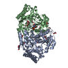
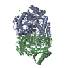
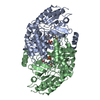
| Title | Structural and functional study of succinyl-ornithine transaminase from E. coli | ||||||
 Components Components | SUCCINYLORNITHINE TRANSAMINASE | ||||||
 Keywords Keywords | TRANSFERASE / PLP ENZYMES / AMINOTRANSFERASE | ||||||
| Function / homology |  Function and homology information Function and homology informationsuccinylornithine transaminase / succinylornithine:2-oxoglutarate transaminase activity / L-ornithine catabolic process / : / N2-acetyl-L-ornithine:2-oxoglutarate 5-transaminase activity / : / : / L-arginine catabolic process / pyridoxal phosphate binding / identical protein binding Similarity search - Function | ||||||
| Biological species |  | ||||||
| Method |  X-RAY DIFFRACTION / SYNCHROTRON / MOLECULAR REPLACEMENT / Resolution: 2.3 Å X-RAY DIFFRACTION / SYNCHROTRON / MOLECULAR REPLACEMENT / Resolution: 2.3 Å | ||||||
 Authors Authors | Newman, J. / Peat, T.S. | ||||||
 Citation Citation | Journal: Plos One / Year: 2013 Title: Determination of the Structure of the Catabolic N-Succinylornithine Transaminase (Astc) from Escherichia Coli. Authors: Newman, J. / Seabrook, S. / Surjadi, R. / Williams, C.C. / Lucent, D. / Wilding, M. / Scott, C. / Peat, T.S. | ||||||
| History |
|
- Structure visualization
| Structure viewer | Molecule: MolmilJmol/JSmol |
|---|
- Downloads & links
-Download
| PDBx/mmCIF format | 4adc.cif.gz | 319.1 KB | Display | PDBx/mmCIF format |
|---|---|---|---|---|
| PDB format | pdb4adc.ent.gz | 260.7 KB | Display | PDB format |
| PDBx/mmJSON format | 4adc.json.gz | Tree view | PDBx/mmJSON format | |
| Others |  Other downloads Other downloads |
-Validation report
| Arichive directory | https://data.pdbj.org/pub/pdb/validation_reports/ad/4adcftp://data.pdbj.org/pub/pdb/validation_reports/ad/4adc | HTTPS FTP |
|---|
-Related structure data
| Related structure data | 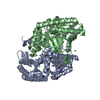 4adbC 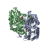 4addC  4adeC  2pb2S C: citing same article ( S: Starting model for refinement |
|---|---|
| Similar structure data |


-Links
 PDBj
PDBj- Assembly
| Deposited unit | 
| |||||||||||||||
|---|---|---|---|---|---|---|---|---|---|---|---|---|---|---|---|---|
| 1 | 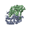
| |||||||||||||||
| 2 | 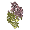
| |||||||||||||||
| Unit cell |
| |||||||||||||||
| Components on special symmetry positions |
|
-Components
| #1: Protein | Mass: 43712.207 Da / Num. of mol.: 4 / Source method: isolated from a natural source / Details: NATIVE FROM E. COLI / Source: (natural) References: UniProt: P77581, succinylornithine transaminase, succinyldiaminopimelate transaminase #2: Chemical | ChemComp-PLP /   Mass: 247.142 Da / Num. of mol.: 4 / Source method: obtained synthetically / Formula: C8H10NO6P Mass: 247.142 Da / Num. of mol.: 4 / Source method: obtained synthetically / Formula: C8H10NO6P#3: Chemical |   Mass: 22.990 Da / Num. of mol.: 2 / Source method: obtained synthetically / Formula: Na Mass: 22.990 Da / Num. of mol.: 2 / Source method: obtained synthetically / Formula: Na#4: Chemical | ChemComp-MG / |   Mass: 24.305 Da / Num. of mol.: 1 / Source method: obtained synthetically / Formula: Mg Mass: 24.305 Da / Num. of mol.: 1 / Source method: obtained synthetically / Formula: Mg#5: Water | ChemComp-HOH / |  Mass: 18.015 Da / Num. of mol.: 518 / Source method: isolated from a natural source / Formula: H2O Mass: 18.015 Da / Num. of mol.: 518 / Source method: isolated from a natural source / Formula: H2ONonpolymer details | PYRIDOXYL PHOSPHATE (PLP): PLP IS FREE FROM LYSINE 252 SCHIFF BASE | |
|---|
-Experimental details
-Experiment
| Experiment | Method: X-RAY DIFFRACTION / Number of used crystals: 1 |
|---|
- Sample preparation
| Crystal | Density Matthews: 3.45 Å3/Da / Density % sol: 64.3 % / Description: NONE |
|---|---|
| Crystal grow | pH: 8 Details: 1.17 M SODIUM MALONATE PH 7.0, 0.09 M TRIS CHLORIDE PH 8.0, 0.02 M DI-ETHYLAMMONIUM FORMATE WITH DRY ORNITHINE POWDER ADDED DIRECTLY TO THE CRYSTAL DROPS. |
-Data collection
| Diffraction | Mean temperature: 100 K |
|---|---|
| Diffraction source | Source: SYNCHROTRON / Site: Australian Synchrotron  / Beamline: MX2 / Wavelength: 0.9537 / Beamline: MX2 / Wavelength: 0.9537 |
| Detector | Type: ADSC QUANTUM 315r / Detector: CCD / Date: Apr 7, 2011 |
| Radiation | Protocol: SINGLE WAVELENGTH / Monochromatic (M) / Laue (L): M / Scattering type: x-ray |
| Radiation wavelength | Wavelength: 0.9537 Å / Relative weight: 1 |
| Reflection | Resolution: 2.3→19.8 Å / Num. obs: 101215 / % possible obs: 97.8 % / Observed criterion σ(I): 1 / Redundancy: 3.5 % / Rmerge(I) obs: 0.1 / Net I/σ(I): 10.7 |
| Reflection shell | Resolution: 2.3→2.42 Å / Redundancy: 2.7 % / Rmerge(I) obs: 0.34 / Mean I/σ(I) obs: 3 / % possible all: 86.6 |
- Processing
| Software |
| ||||||||||||||||||||||||||||||||||||||||||||||||||||||||||||||||||||||||||||||||||||||||||||||||||||||||||||||||||||||||||||||||||||||||||||||||||||||||||||||||||||||||||||||||||||||
|---|---|---|---|---|---|---|---|---|---|---|---|---|---|---|---|---|---|---|---|---|---|---|---|---|---|---|---|---|---|---|---|---|---|---|---|---|---|---|---|---|---|---|---|---|---|---|---|---|---|---|---|---|---|---|---|---|---|---|---|---|---|---|---|---|---|---|---|---|---|---|---|---|---|---|---|---|---|---|---|---|---|---|---|---|---|---|---|---|---|---|---|---|---|---|---|---|---|---|---|---|---|---|---|---|---|---|---|---|---|---|---|---|---|---|---|---|---|---|---|---|---|---|---|---|---|---|---|---|---|---|---|---|---|---|---|---|---|---|---|---|---|---|---|---|---|---|---|---|---|---|---|---|---|---|---|---|---|---|---|---|---|---|---|---|---|---|---|---|---|---|---|---|---|---|---|---|---|---|---|---|---|---|---|
| Refinement | Method to determine structure: MOLECULAR REPLACEMENT Starting model: PDB ENTRY 2PB2 Resolution: 2.3→108.69 Å / Cor.coef. Fo:Fc: 0.95 / Cor.coef. Fo:Fc free: 0.922 / SU B: 5.582 / SU ML: 0.13 / Cross valid method: THROUGHOUT / ESU R: 0.216 / ESU R Free: 0.184 / Stereochemistry target values: MAXIMUM LIKELIHOOD Details: HYDROGENS HAVE BEEN USED IF PRESENT IN THE INPUT. U VALUES REFINED INDIVIDUALLY.
| ||||||||||||||||||||||||||||||||||||||||||||||||||||||||||||||||||||||||||||||||||||||||||||||||||||||||||||||||||||||||||||||||||||||||||||||||||||||||||||||||||||||||||||||||||||||
| Solvent computation | Ion probe radii: 0.8 Å / Shrinkage radii: 0.8 Å / VDW probe radii: 1.2 Å / Solvent model: MASK | ||||||||||||||||||||||||||||||||||||||||||||||||||||||||||||||||||||||||||||||||||||||||||||||||||||||||||||||||||||||||||||||||||||||||||||||||||||||||||||||||||||||||||||||||||||||
| Displacement parameters | Biso mean: 23.519 Å2
| ||||||||||||||||||||||||||||||||||||||||||||||||||||||||||||||||||||||||||||||||||||||||||||||||||||||||||||||||||||||||||||||||||||||||||||||||||||||||||||||||||||||||||||||||||||||
| Refinement step | Cycle: LAST / Resolution: 2.3→108.69 Å
| ||||||||||||||||||||||||||||||||||||||||||||||||||||||||||||||||||||||||||||||||||||||||||||||||||||||||||||||||||||||||||||||||||||||||||||||||||||||||||||||||||||||||||||||||||||||
| Refine LS restraints |
|