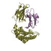
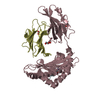
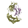
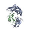
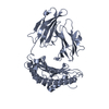
[English] 日本語
Yorodumi
- PDB-4aco: Structure of the budding yeast Ndc10 N-terminal domain -

+
Open data


ID or keywords:

Loading...

-
Basic information

Entry
Database: PDB / ID: 4aco
TitleStructure of the budding yeast Ndc10 N-terminal domain
ComponentsCENTROMERE DNA-BINDING PROTEIN COMPLEX CBF3 SUBUNIT A
KeywordsDNA BINDING PROTEIN
Function / homology
Function and homology information


CBF3 complex / septin ring assembly / mitotic spindle elongation / centromeric DNA binding / kinetochore assembly / condensed chromosome, centromeric region / spindle pole body / DNA binding, bending / spindle midzone / chromosome segregation ...CBF3 complex / septin ring assembly / mitotic spindle elongation / centromeric DNA binding / kinetochore assembly / condensed chromosome, centromeric region / spindle pole body / DNA binding, bending / spindle midzone / chromosome segregation / spindle / kinetochore / DNA binding / nucleus / cytoplasm
Similarity search - Function
Centromere DNA-binding protein complex CBF3 subunit, domain 2 / : / Ndc10 N-terminal domain / Transcription activator GCR1-like domain / Ndc10, domain 2 / Ndc10, domain 2 superfamily / Transcriptional activator of glycolytic enzymes / Centromere DNA-binding protein complex CBF3 subunit, domain 2 / Tyrosine recombinase, N-terminal domain / hpI Integrase; Chain A ...Centromere DNA-binding protein complex CBF3 subunit, domain 2 / : / Ndc10 N-terminal domain / Transcription activator GCR1-like domain / Ndc10, domain 2 / Ndc10, domain 2 superfamily / Transcriptional activator of glycolytic enzymes / Centromere DNA-binding protein complex CBF3 subunit, domain 2 / Tyrosine recombinase, N-terminal domain / hpI Integrase; Chain A / Integrase/recombinase, N-terminal / DNA polymerase; domain 1 / Orthogonal Bundle / Mainly Alpha
Similarity search - Domain/homology
Centromere DNA-binding protein complex CBF3 subunit A
Similarity search - Component
Biological speciesSACCHAROMYCES CEREVISIAE (brewer's yeast)
MethodX-RAY DIFFRACTION / SYNCHROTRON / OTHER / Resolution: 1.89 Å
AuthorsPerriches, T. / Singleton, M.R.
CitationJournal: J.Biol.Chem. / Year: 2012
Title: The Structure of the Yeast Kinetochore Ndc10 DNA-Binding Domain Reveals an Unexpected Evolutionary Relationship to Tyrosine Recombinases.
Authors: Perriches, T. / Singleton, M.R.
History
DepositionDec 16, 2011Deposition site: PDBE / Processing site: PDBE
Revision 1.0Jan 11, 2012Provider: repository / Type: Initial release
Revision 1.1Jan 25, 2012Group: Other
Revision 1.2Feb 29, 2012Group: Other

-
Structure visualization

Structure viewerMolecule:
MolmilJmol/JSmol

Downloads & links

-
Assembly

Deposited unit
A: CENTROMERE DNA-BINDING PROTEIN COMPLEX CBF3 SUBUNIT A


Theoretical massNumber of molelcules
Total (without water)112,0661
Polymers112,0661
Non-polymers00
Water1,54986
1


  • Idetical with deposited unit
  • defined by author&software
TypeNameSymmetry operationNumber
identity operation1_555x,y,z1
MethodPISA
Unit cell
Length a, b, c (Å)55.740, 87.760, 104.570
Angle α, β, γ (deg.)90.00, 90.00, 90.00
Int Tables number19
Space group name H-MP212121

-
Components

#1: Protein CENTROMERE DNA-BINDING PROTEIN COMPLEX CBF3 SUBUNIT A / CENTROMERE-BINDING FACTOR 2 / CHROMOSOME TRANSMISSION FIDELITY PROTEIN 14 / KINETOCHORE PROTEIN CTF14 / NDC10


Mass: 112066.031 Da / Num. of mol.: 1
Source method: isolated from a genetically manipulated source
Source: (gene. exp.) SACCHAROMYCES CEREVISIAE (brewer's yeast)
Production host: ESCHERICHIA COLI (E. coli) / References: UniProt: P32504
#2: Water ChemComp-HOH / water / Water


Mass: 18.015 Da / Num. of mol.: 86 / Source method: isolated from a natural source / Formula: H2O

-
Experimental details

-
Experiment

ExperimentMethod: X-RAY DIFFRACTION

-
Sample preparation

CrystalDensity Matthews: 2.35 Å3/Da / Density % sol: 47.36 % / Description: NONE

-
Data collection

DiffractionMean temperature: 100 K
Diffraction sourceSource: SYNCHROTRON / Site: Diamond / Beamline: I24 / Wavelength: 0.97
DetectorType: ADSC CCD / Detector: CCD
RadiationProtocol: SINGLE WAVELENGTH / Monochromatic (M) / Laue (L): M / Scattering type: x-ray
Radiation wavelengthWavelength: 0.97 Å / Relative weight: 1
ReflectionResolution: 1.89→67.22 Å / Num. obs: 39129 / % possible obs: 98.4 % / Redundancy: 3.3 % / Rmerge(I) obs: 0.05 / Net I/σ(I): 13.4

-
Processing

SoftwareName: REFMAC / Version: 5.6.0117 / Classification: refinement
RefinementMethod to determine structure: OTHER
Starting model: NONE

Resolution: 1.89→67.22 Å / Cor.coef. Fo:Fc: 0.953 / Cor.coef. Fo:Fc free: 0.936 / SU B: 3.147 / SU ML: 0.093 / Cross valid method: THROUGHOUT / ESU R: 0.151 / ESU R Free: 0.141 / Stereochemistry target values: MAXIMUM LIKELIHOOD / Details: HYDROGENS HAVE BEEN ADDED IN THE RIDING POSITIONS.
RfactorNum. reflection% reflectionSelection details
Rfree0.23159 2054 5 %RANDOM
Rwork0.19253 ---
obs0.19443 39129 98.42 %-
Solvent computationIon probe radii: 0.8 Å / Shrinkage radii: 0.8 Å / VDW probe radii: 1.2 Å / Solvent model: MASK
Displacement parametersBiso mean: 28.123 Å2
Baniso -1Baniso -2Baniso -3
1-0.56 Å20 Å20 Å2
2---0.49 Å20 Å2
3----0.07 Å2
Refinement stepCycle: LAST / Resolution: 1.89→67.22 Å
ProteinNucleic acidLigandSolventTotal
Num. atoms3793 0 0 86 3879
Refine LS restraints
Refine-IDTypeDev idealDev ideal targetNumber
X-RAY DIFFRACTIONr_bond_refined_d0.020.023908
X-RAY DIFFRACTIONr_bond_other_d
X-RAY DIFFRACTIONr_angle_refined_deg2.0341.9585297
X-RAY DIFFRACTIONr_angle_other_deg
X-RAY DIFFRACTIONr_dihedral_angle_1_deg6.1685446
X-RAY DIFFRACTIONr_dihedral_angle_2_deg37.20824.129201
X-RAY DIFFRACTIONr_dihedral_angle_3_deg18.13615666
X-RAY DIFFRACTIONr_dihedral_angle_4_deg15.8241518
X-RAY DIFFRACTIONr_chiral_restr0.1510.2557
X-RAY DIFFRACTIONr_gen_planes_refined0.0120.0213008
X-RAY DIFFRACTIONr_gen_planes_other
X-RAY DIFFRACTIONr_nbd_refined
X-RAY DIFFRACTIONr_nbd_other
X-RAY DIFFRACTIONr_nbtor_refined
X-RAY DIFFRACTIONr_nbtor_other
X-RAY DIFFRACTIONr_xyhbond_nbd_refined
X-RAY DIFFRACTIONr_xyhbond_nbd_other
X-RAY DIFFRACTIONr_metal_ion_refined
X-RAY DIFFRACTIONr_metal_ion_other
X-RAY DIFFRACTIONr_symmetry_vdw_refined
X-RAY DIFFRACTIONr_symmetry_vdw_other
X-RAY DIFFRACTIONr_symmetry_hbond_refined
X-RAY DIFFRACTIONr_symmetry_hbond_other
X-RAY DIFFRACTIONr_symmetry_metal_ion_refined
X-RAY DIFFRACTIONr_symmetry_metal_ion_other
X-RAY DIFFRACTIONr_mcbond_it
X-RAY DIFFRACTIONr_mcbond_other
X-RAY DIFFRACTIONr_mcangle_it
X-RAY DIFFRACTIONr_mcangle_other
X-RAY DIFFRACTIONr_scbond_it
X-RAY DIFFRACTIONr_scbond_other
X-RAY DIFFRACTIONr_scangle_it
X-RAY DIFFRACTIONr_scangle_other
X-RAY DIFFRACTIONr_long_range_B_refined
X-RAY DIFFRACTIONr_long_range_B_other
X-RAY DIFFRACTIONr_rigid_bond_restr
X-RAY DIFFRACTIONr_sphericity_free
X-RAY DIFFRACTIONr_sphericity_bonded
LS refinement shellResolution: 1.89→1.939 Å / Total num. of bins used: 20
RfactorNum. reflection% reflection
Rfree0.354 157 -
Rwork0.29 2637 -
obs--98.41 %

+
About Yorodumi

-
News

-
Feb 9, 2022. New format data for meta-information of EMDB entries

New format data for meta-information of EMDB entries

  • Version 3 of the EMDB header file is now the official format.
  • The previous official version 1.9 will be removed from the archive.

Related info.:EMDB header

External links:wwPDB to switch to version 3 of the EMDB data model

-
Aug 12, 2020. Covid-19 info

Covid-19 info

URL: https://pdbj.org/emnavi/covid19.php

New page: Covid-19 featured information page in EM Navigator.

Related info.:Covid-19 info / Mar 5, 2020. Novel coronavirus structure data

+
Mar 5, 2020. Novel coronavirus structure data

Novel coronavirus structure data

Related info.:Yorodumi Speices / Aug 12, 2020. Covid-19 info

External links:COVID-19 featured content - PDBj / Molecule of the Month (242):Coronavirus Proteases

+
Jan 31, 2019. EMDB accession codes are about to change! (news from PDBe EMDB page)

EMDB accession codes are about to change! (news from PDBe EMDB page)

  • The allocation of 4 digits for EMDB accession codes will soon come to an end. Whilst these codes will remain in use, new EMDB accession codes will include an additional digit and will expand incrementally as the available range of codes is exhausted. The current 4-digit format prefixed with “EMD-” (i.e. EMD-XXXX) will advance to a 5-digit format (i.e. EMD-XXXXX), and so on. It is currently estimated that the 4-digit codes will be depleted around Spring 2019, at which point the 5-digit format will come into force.
  • The EM Navigator/Yorodumi systems omit the EMD- prefix.

Related info.:Q: What is EMD? / ID/Accession-code notation in Yorodumi/EM Navigator

External links:EMDB Accession Codes are Changing Soon! / Contact to PDBj

+
Jul 12, 2017. Major update of PDB

Major update of PDB

  • wwPDB released updated PDB data conforming to the new PDBx/mmCIF dictionary.
  • This is a major update changing the version number from 4 to 5, and with Remediation, in which all the entries are updated.
  • In this update, many items about electron microscopy experimental information are reorganized (e.g. em_software).
  • Now, EM Navigator and Yorodumi are based on the updated data.

External links:wwPDB Remediation / Enriched Model Files Conforming to OneDep Data Standards Now Available in the PDB FTP Archive

-
Yorodumi

Thousand views of thousand structures

  • Yorodumi is a browser for structure data from EMDB, PDB, SASBDB, etc.
  • This page is also the successor to EM Navigator detail page, and also detail information page/front-end page for Omokage search.
  • The word "yorodu" (or yorozu) is an old Japanese word meaning "ten thousand". "mi" (miru) is to see.

Related info.:EMDB / PDB / SASBDB / Comparison of 3 databanks / Yorodumi Search / Aug 31, 2016. New EM Navigator & Yorodumi / Yorodumi Papers / Jmol/JSmol / Function and homology information / Changes in new EM Navigator and Yorodumi

Read more