 Movie
Movie Controller

[English] 日本語
 Yorodumi
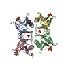
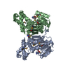
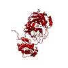
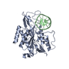
- PDB-4a6f: Crystal structure of Slm1-PH domain in complex with Phosphoserine -
+ Open data

- Basic information
| Entry | Database: PDB / ID: 4a6f | ||||||
|---|---|---|---|---|---|---|---|
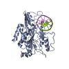
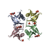
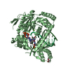
| Title | Crystal structure of Slm1-PH domain in complex with Phosphoserine | ||||||
 Components Components | (PHOSPHATIDYLINOSITOL 4,5-BISPHOSPHATE-BINDING PROTEIN SLM1) x 2 | ||||||
 Keywords Keywords | SIGNALING PROTEIN / POST TRANSLATIONAL MODIFICATION | ||||||
| Function / homology |  Function and homology information Function and homology informationeisosome assembly / sphingolipid binding / establishment or maintenance of actin cytoskeleton polarity / sphingolipid metabolic process / endosomal transport / actin filament bundle assembly / TOR signaling / phosphatidylinositol-4,5-bisphosphate binding / protein localization to plasma membrane / regulation of cell growth ...eisosome assembly / sphingolipid binding / establishment or maintenance of actin cytoskeleton polarity / sphingolipid metabolic process / endosomal transport / actin filament bundle assembly / TOR signaling / phosphatidylinositol-4,5-bisphosphate binding / protein localization to plasma membrane / regulation of cell growth / actin cytoskeleton organization / mitochondrion / identical protein binding / plasma membrane / cytoplasm Similarity search - Function | ||||||
| Biological species |  | ||||||
| Method |  X-RAY DIFFRACTION / SYNCHROTRON / MOLECULAR REPLACEMENT / Resolution: 1.68 Å X-RAY DIFFRACTION / SYNCHROTRON / MOLECULAR REPLACEMENT / Resolution: 1.68 Å | ||||||
 Authors Authors | Anand, K. / Maeda, K. / Gavin, A.C. | ||||||
 Citation Citation | Journal: Plos One / Year: 2012 Title: Structural Analyses of Slm1-Ph Domain Demonstrate Ligand Binding in the Non-Canonical Site Authors: Anand, K. / Maeda, K. / Gavin, A.C. | ||||||
| History |
|
- Structure visualization
| Structure viewer | Molecule: MolmilJmol/JSmol |
|---|
- Downloads & links
-Download
| PDBx/mmCIF format | 4a6f.cif.gz | 106.2 KB | Display | PDBx/mmCIF format |
|---|---|---|---|---|
| PDB format | pdb4a6f.ent.gz | 80.9 KB | Display | PDB format |
| PDBx/mmJSON format | 4a6f.json.gz | Tree view | PDBx/mmJSON format | |
| Others |  Other downloads Other downloads |
-Validation report
| Arichive directory | https://data.pdbj.org/pub/pdb/validation_reports/a6/4a6fftp://data.pdbj.org/pub/pdb/validation_reports/a6/4a6f | HTTPS FTP |
|---|
-Related structure data
| Related structure data | 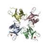 4a5kSC  4a6hC  4a6kC S: Starting model for refinement C: citing same article ( |
|---|---|
| Similar structure data |


-Links
 PDBj
PDBj

- Assembly
| Deposited unit | 
| |||||||||
|---|---|---|---|---|---|---|---|---|---|---|
| 1 | 
| |||||||||
| Unit cell |
| |||||||||
| Components on special symmetry positions |
|
-Components
| #1: Protein | Mass: 13777.617 Da / Num. of mol.: 1 / Fragment: SLM1-PH DOMAIN, RESIDUES 469-583 Source method: isolated from a genetically manipulated source Source: (gene. exp.) Production host:  | ||||
|---|---|---|---|---|---|
| #2: Protein | Mass: 13754.582 Da / Num. of mol.: 1 / Fragment: SLM1-PH DOMAIN, RESIDUES 469-583 Source method: isolated from a genetically manipulated source Source: (gene. exp.) Production host: | ||||
| #3: Chemical |   Mass: 94.971 Da / Num. of mol.: 2 / Source method: obtained synthetically / Formula: PO4 Mass: 94.971 Da / Num. of mol.: 2 / Source method: obtained synthetically / Formula: PO4#4: Chemical | ChemComp-SEP /   Type: L-peptide linking / Mass: 185.072 Da / Num. of mol.: 4 / Source method: obtained synthetically / Formula: C3H8NO6P Type: L-peptide linking / Mass: 185.072 Da / Num. of mol.: 4 / Source method: obtained synthetically / Formula: C3H8NO6P#5: Water | ChemComp-HOH / |  Mass: 18.015 Da / Num. of mol.: 174 / Source method: isolated from a natural source / Formula: H2O Mass: 18.015 Da / Num. of mol.: 174 / Source method: isolated from a natural source / Formula: H2O |
-Experimental details
-Experiment
| Experiment | Method: X-RAY DIFFRACTION / Number of used crystals: 1 |
|---|
- Sample preparation
| Crystal | Density Matthews: 2.2 Å3/Da / Density % sol: 44 % / Description: NONE |
|---|---|
| Crystal grow | pH: 7.5 / Details: pH 7.5 |
-Data collection
| Diffraction | Mean temperature: 100 K |
|---|---|
| Diffraction source | Source: SYNCHROTRON / Site: SLS  / Beamline: X06DA / Wavelength: 1 / Beamline: X06DA / Wavelength: 1 |
| Detector | Type: MARRESEARCH / Detector: CCD / Date: Oct 9, 2009 / Details: MIRRORS |
| Radiation | Protocol: SINGLE WAVELENGTH / Monochromatic (M) / Laue (L): M / Scattering type: x-ray |
| Radiation wavelength | Wavelength: 1 Å / Relative weight: 1 |
| Reflection | Resolution: 1.68→20 Å / Num. obs: 26485 / % possible obs: 98.7 % / Observed criterion σ(I): 2.34 / Redundancy: 5.4 % / Rmerge(I) obs: 0.1 / Net I/σ(I): 10.61 |
| Reflection shell | Resolution: 1.68→1.8 Å / Redundancy: 3.6 % / Rmerge(I) obs: 0.5 / Mean I/σ(I) obs: 2.34 / % possible all: 93.4 |
- Processing
| Software |
| |||||||||||||||||||||||||||||||||||||||||||||||||||||||||||||||
|---|---|---|---|---|---|---|---|---|---|---|---|---|---|---|---|---|---|---|---|---|---|---|---|---|---|---|---|---|---|---|---|---|---|---|---|---|---|---|---|---|---|---|---|---|---|---|---|---|---|---|---|---|---|---|---|---|---|---|---|---|---|---|---|---|
| Refinement | Method to determine structure: MOLECULAR REPLACEMENT Starting model: PDB ENTRY 4A5K Resolution: 1.68→54.933 Å / SU ML: 0.21 / σ(F): 2.02 / Phase error: 23.34 / Stereochemistry target values: ML
| |||||||||||||||||||||||||||||||||||||||||||||||||||||||||||||||
| Solvent computation | Shrinkage radii: 0.9 Å / VDW probe radii: 1.11 Å / Solvent model: FLAT BULK SOLVENT MODEL / Bsol: 43.803 Å2 / ksol: 0.357 e/Å3 | |||||||||||||||||||||||||||||||||||||||||||||||||||||||||||||||
| Displacement parameters |
| |||||||||||||||||||||||||||||||||||||||||||||||||||||||||||||||
| Refinement step | Cycle: LAST / Resolution: 1.68→54.933 Å
| |||||||||||||||||||||||||||||||||||||||||||||||||||||||||||||||
| Refine LS restraints |
| |||||||||||||||||||||||||||||||||||||||||||||||||||||||||||||||
| LS refinement shell |
| |||||||||||||||||||||||||||||||||||||||||||||||||||||||||||||||
| Refinement TLS params. | Method: refined / Origin x: 0.3378 Å / Origin y: -7.506 Å / Origin z: 9.3801 Å
| |||||||||||||||||||||||||||||||||||||||||||||||||||||||||||||||
| Refinement TLS group | Selection details: ALL |
