 Movie
Movie Controller

[English] 日本語
 Yorodumi
- PDB-4a2o: STRUCTURE OF THE HUMAN EOSINOPHIL CATIONIC PROTEIN IN COMPLEX WIT... -
+ Open data

- Basic information
| Entry | Database: PDB / ID: 4a2o | ||||||
|---|---|---|---|---|---|---|---|
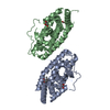
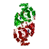
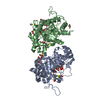
| Title | STRUCTURE OF THE HUMAN EOSINOPHIL CATIONIC PROTEIN IN COMPLEX WITH SULFATE ANIONS | ||||||
 Components Components | EOSINOPHIL CATIONIC PROTEIN | ||||||
 Keywords Keywords | HYDROLASE / OXIDOREDUCTASE / ANTIMICROBIAL / CYTOTOXIC | ||||||
| Function / homology |  Function and homology information Function and homology informationinduction of bacterial agglutination / Hydrolases; Acting on ester bonds; Endoribonucleases producing 3'-phosphomonoesters / RNA catabolic process / Antimicrobial peptides / RNA nuclease activity / innate immune response in mucosa / lipopolysaccharide binding / chemotaxis / azurophil granule lumen / antimicrobial humoral immune response mediated by antimicrobial peptide ...induction of bacterial agglutination / Hydrolases; Acting on ester bonds; Endoribonucleases producing 3'-phosphomonoesters / RNA catabolic process / Antimicrobial peptides / RNA nuclease activity / innate immune response in mucosa / lipopolysaccharide binding / chemotaxis / azurophil granule lumen / antimicrobial humoral immune response mediated by antimicrobial peptide / antibacterial humoral response / endonuclease activity / defense response to Gram-negative bacterium / nucleic acid binding / defense response to Gram-positive bacterium / innate immune response / hydrolase activity / Neutrophil degranulation / : / extracellular region Similarity search - Function | ||||||
| Biological species |  HOMO SAPIENS (human) HOMO SAPIENS (human) | ||||||
| Method |  X-RAY DIFFRACTION / SYNCHROTRON / MOLECULAR REPLACEMENT / Resolution: 1.69 Å X-RAY DIFFRACTION / SYNCHROTRON / MOLECULAR REPLACEMENT / Resolution: 1.69 Å | ||||||
 Authors Authors | Boix, E. / Pulido, D. / Moussaoui, M. / Nogues, V. / Russi, S. | ||||||
 Citation Citation | Journal: J.Struct.Biol. / Year: 2012 Title: The Sulfate-Binding Site Structure of the Human Eosinophil Cationic Protein as Revealed by a New Crystal Form. Authors: Boix, E. / Pulido, D. / Moussaoui, M. / Nogues, V. / Russi, S. | ||||||
| History |
|
- Structure visualization
| Structure viewer | Molecule: MolmilJmol/JSmol |
|---|
- Downloads & links
-Download
| PDBx/mmCIF format | 4a2o.cif.gz | 78.2 KB | Display | PDBx/mmCIF format |
|---|---|---|---|---|
| PDB format | pdb4a2o.ent.gz | 59.5 KB | Display | PDB format |
| PDBx/mmJSON format | 4a2o.json.gz | Tree view | PDBx/mmJSON format | |
| Others |  Other downloads Other downloads |
-Validation report
| Arichive directory | https://data.pdbj.org/pub/pdb/validation_reports/a2/4a2oftp://data.pdbj.org/pub/pdb/validation_reports/a2/4a2o | HTTPS FTP |
|---|
-Related structure data
| Related structure data |  4a2yC  1dytS C: citing same article ( S: Starting model for refinement |
|---|---|
| Similar structure data |


-Links
 PDBj
PDBj


- Assembly
| Deposited unit | 
| ||||||||
|---|---|---|---|---|---|---|---|---|---|
| 1 | 
| ||||||||
| Unit cell |
| ||||||||
| Components on special symmetry positions |
|
-Components
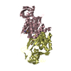
| #1: Protein | Mass: 15598.876 Da / Num. of mol.: 2 Source method: isolated from a genetically manipulated source Source: (gene. exp.) HOMO SAPIENS (human) / Cell: EOSINOPHIL / Plasmid: PET11C/ECP / Production host:  References: UniProt: P12724, Hydrolases; Acting on ester bonds; Endoribonucleases producing 3'-phosphomonoesters #2: Chemical | ChemComp-SO4 /   Mass: 96.063 Da / Num. of mol.: 14 / Source method: obtained synthetically / Formula: SO4 Mass: 96.063 Da / Num. of mol.: 14 / Source method: obtained synthetically / Formula: SO4#3: Water | ChemComp-HOH / |  Mass: 18.015 Da / Num. of mol.: 371 / Source method: isolated from a natural source / Formula: H2O Mass: 18.015 Da / Num. of mol.: 371 / Source method: isolated from a natural source / Formula: H2OHas protein modification | Y | Sequence details | RECOMBINANT PROTEIN EXPRESSED USING A SYNTHETIC GENE (BOIX ET AL. J.BIOL.CHEM. 274, 15605) BASED ON ...RECOMBINAN | |
|---|
-Experimental details
-Experiment
| Experiment | Method: X-RAY DIFFRACTION / Number of used crystals: 1 |
|---|
- Sample preparation
| Crystal | Density Matthews: 1.63 Å3/Da / Density % sol: 24.17 % / Description: NONE |
|---|---|
| Crystal grow | pH: 8.5 / Details: pH 8.5 |
-Data collection
| Diffraction | Mean temperature: 100 K |
|---|---|
| Diffraction source | Source: SYNCHROTRON / Site: ESRF  / Beamline: BM14 / Wavelength: 0.9537 / Beamline: BM14 / Wavelength: 0.9537 |
| Detector | Type: MARMOSAIC 225 mm CCD / Detector: CCD / Date: Jul 1, 2011 |
| Radiation | Protocol: SINGLE WAVELENGTH / Monochromatic (M) / Laue (L): M / Scattering type: x-ray |
| Radiation wavelength | Wavelength: 0.9537 Å / Relative weight: 1 |
| Reflection | Resolution: 1.69→51.62 Å / Num. obs: 25341 / % possible obs: 98.5 % / Observed criterion σ(I): 2 / Redundancy: 3 % / Biso Wilson estimate: 23.6 Å2 / Rmerge(I) obs: 0.07 / Net I/σ(I): 11.4 |
| Reflection shell | Resolution: 1.69→1.78 Å / Redundancy: 2.3 % / Rmerge(I) obs: 0.45 / Mean I/σ(I) obs: 2.3 / % possible all: 90.8 |
- Processing
| Software |
| ||||||||||||||||||||||||||||||||||||||||||||||||||||||||||||||||||||||||||||||||||||||||||||||||||||||||||||||||||||||||||||||||||||||||||||||||||||||||||||||||||||||||||||||||||||||
|---|---|---|---|---|---|---|---|---|---|---|---|---|---|---|---|---|---|---|---|---|---|---|---|---|---|---|---|---|---|---|---|---|---|---|---|---|---|---|---|---|---|---|---|---|---|---|---|---|---|---|---|---|---|---|---|---|---|---|---|---|---|---|---|---|---|---|---|---|---|---|---|---|---|---|---|---|---|---|---|---|---|---|---|---|---|---|---|---|---|---|---|---|---|---|---|---|---|---|---|---|---|---|---|---|---|---|---|---|---|---|---|---|---|---|---|---|---|---|---|---|---|---|---|---|---|---|---|---|---|---|---|---|---|---|---|---|---|---|---|---|---|---|---|---|---|---|---|---|---|---|---|---|---|---|---|---|---|---|---|---|---|---|---|---|---|---|---|---|---|---|---|---|---|---|---|---|---|---|---|---|---|---|---|
| Refinement | Method to determine structure: MOLECULAR REPLACEMENT Starting model: PDB ENTRY 1DYT Resolution: 1.69→51.62 Å / Cor.coef. Fo:Fc: 0.961 / Cor.coef. Fo:Fc free: 0.929 / SU B: 2.42 / SU ML: 0.081 / Cross valid method: THROUGHOUT / ESU R: 0.12 / ESU R Free: 0.125 / Stereochemistry target values: MAXIMUM LIKELIHOOD Details: HYDROGENS HAVE BEEN ADDED IN THE RIDING POSITIONS. RESIDUES 1-27 OF P12724 ARE THE SIGNAL PEPTIDE. LOOP 90-92 IS PARTIALLY DISORDERED SIDE CHAINS OF R28, W35, R73, F76, R101, AND R105 ARE PARTIALLY DISORDERED.
| ||||||||||||||||||||||||||||||||||||||||||||||||||||||||||||||||||||||||||||||||||||||||||||||||||||||||||||||||||||||||||||||||||||||||||||||||||||||||||||||||||||||||||||||||||||||
| Solvent computation | Ion probe radii: 0.8 Å / Shrinkage radii: 0.8 Å / VDW probe radii: 1.4 Å / Solvent model: MASK | ||||||||||||||||||||||||||||||||||||||||||||||||||||||||||||||||||||||||||||||||||||||||||||||||||||||||||||||||||||||||||||||||||||||||||||||||||||||||||||||||||||||||||||||||||||||
| Displacement parameters | Biso mean: 16.866 Å2
| ||||||||||||||||||||||||||||||||||||||||||||||||||||||||||||||||||||||||||||||||||||||||||||||||||||||||||||||||||||||||||||||||||||||||||||||||||||||||||||||||||||||||||||||||||||||
| Refinement step | Cycle: LAST / Resolution: 1.69→51.62 Å
| ||||||||||||||||||||||||||||||||||||||||||||||||||||||||||||||||||||||||||||||||||||||||||||||||||||||||||||||||||||||||||||||||||||||||||||||||||||||||||||||||||||||||||||||||||||||
| Refine LS restraints |
|