 Movie
Movie Controller

[English] 日本語
 Yorodumi
- PDB-1h1h: Crystal Structure of Eosinophil Cationic Protein in Complex with ... -
+ Open data

- Basic information
| Entry | Database: PDB / ID: 1h1h | |||||||||
|---|---|---|---|---|---|---|---|---|---|---|
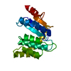
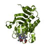
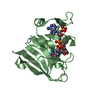
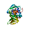
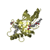
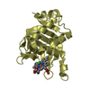
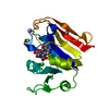
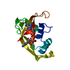
| Title | Crystal Structure of Eosinophil Cationic Protein in Complex with 2',5'-ADP at 2.0 A resolution Reveals the Details of the Ribonucleolytic Active site | |||||||||
 Components Components | EOSINOPHIL CATIONIC PROTEIN | |||||||||
 Keywords Keywords | HYDROLASE / EOSINOPHIL CATIONIC PROTEIN / EOSINOPHIL DERIVED NEUROTOXIN / ADENOSINE-2' / 5'-DIPHOSPHATE / RIBONUCLEASE / TOXIN | |||||||||
| Function / homology |  Function and homology information Function and homology informationinduction of bacterial agglutination / Hydrolases; Acting on ester bonds; Endoribonucleases producing 3'-phosphomonoesters / RNA catabolic process / Antimicrobial peptides / RNA nuclease activity / innate immune response in mucosa / lipopolysaccharide binding / chemotaxis / azurophil granule lumen / antimicrobial humoral immune response mediated by antimicrobial peptide ...induction of bacterial agglutination / Hydrolases; Acting on ester bonds; Endoribonucleases producing 3'-phosphomonoesters / RNA catabolic process / Antimicrobial peptides / RNA nuclease activity / innate immune response in mucosa / lipopolysaccharide binding / chemotaxis / azurophil granule lumen / antimicrobial humoral immune response mediated by antimicrobial peptide / antibacterial humoral response / endonuclease activity / nucleic acid binding / defense response to Gram-negative bacterium / defense response to Gram-positive bacterium / innate immune response / hydrolase activity / Neutrophil degranulation / : / extracellular region Similarity search - Function | |||||||||
| Biological species |  HOMO SAPIENS (human) HOMO SAPIENS (human) | |||||||||
| Method |  X-RAY DIFFRACTION / SYNCHROTRON / MOLECULAR REPLACEMENT / Resolution: 2 Å X-RAY DIFFRACTION / SYNCHROTRON / MOLECULAR REPLACEMENT / Resolution: 2 Å | |||||||||
 Authors Authors | Mohan, C.G. / Boix, E. / Evans, H.R. / Nikolovski, Z. / Nogues, M.V. / Cuchillo, C.M. / Acharya, K.R. | |||||||||
 Citation Citation | Journal: Biochemistry / Year: 2002 Title: The Crystal Structure of Eosinophil Cationic Protein in Complex with 2'5'-Adp at 2.0 A Resolution Reveals the Details of the Ribonucleolytic Active Site Authors: Mohan, C.G. / Boix, E. / Evans, H.R. / Nikolovski, Z. / Nogues, M.V. / Cuchillo, C.M. / Acharya, K.R. | |||||||||
| History |
| |||||||||
| Remark 700 | SHEET THE SHEET STRUCTURE OF THIS MOLECULE IS BIFURCATED. IN ORDER TO REPRESENT THIS FEATURE IN ... SHEET THE SHEET STRUCTURE OF THIS MOLECULE IS BIFURCATED. IN ORDER TO REPRESENT THIS FEATURE IN THE SHEET RECORDS BELOW, TWO SHEETS ARE DEFINED. |
- Structure visualization
| Structure viewer | Molecule: MolmilJmol/JSmol |
|---|
- Downloads & links
-Download
| PDBx/mmCIF format | 1h1h.cif.gz | 42.7 KB | Display | PDBx/mmCIF format |
|---|---|---|---|---|
| PDB format | pdb1h1h.ent.gz | 29.5 KB | Display | PDB format |
| PDBx/mmJSON format | 1h1h.json.gz | Tree view | PDBx/mmJSON format | |
| Others |  Other downloads Other downloads |
-Validation report
| Arichive directory | https://data.pdbj.org/pub/pdb/validation_reports/h1/1h1hftp://data.pdbj.org/pub/pdb/validation_reports/h1/1h1h | HTTPS FTP |
|---|
-Related structure data
| Related structure data |  1qmtS S: Starting model for refinement |
|---|---|
| Similar structure data |


-Links
 PDBj
PDBj


- Assembly
| Deposited unit | 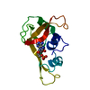
| ||||||||
|---|---|---|---|---|---|---|---|---|---|
| 1 |
| ||||||||
| Unit cell |
|
-Components
| #1: Protein | Mass: 15730.072 Da / Num. of mol.: 1 Source method: isolated from a genetically manipulated source Source: (gene. exp.) HOMO SAPIENS (human) / Production host:  References: UniProt: P12724, Hydrolases; Acting on ester bonds; Endoribonucleases producing 3'-phosphomonoesters | ||
|---|---|---|---|
| #2: Chemical | ChemComp-A2P /   Mass: 427.201 Da / Num. of mol.: 1 / Source method: obtained synthetically / Formula: C10H15N5O10P2 Mass: 427.201 Da / Num. of mol.: 1 / Source method: obtained synthetically / Formula: C10H15N5O10P2 | ||
| #3: Water | ChemComp-HOH /  Mass: 18.015 Da / Num. of mol.: 54 / Source method: isolated from a natural source / Formula: H2O Mass: 18.015 Da / Num. of mol.: 54 / Source method: isolated from a natural source / Formula: H2O | ||
| Compound details | CYTOTOXIN & HELMINTHOT| Has protein modification | Y | |
-Experimental details
-Experiment
| Experiment | Method: X-RAY DIFFRACTION / Number of used crystals: 1 |
|---|
- Sample preparation
| Crystal | Density Matthews: 2.95 Å3/Da / Density % sol: 58.3 % | ||||||||||||||||||||||||||||||
|---|---|---|---|---|---|---|---|---|---|---|---|---|---|---|---|---|---|---|---|---|---|---|---|---|---|---|---|---|---|---|---|
| Crystal grow | pH: 8 Details: 0.5 M SODIUM CHLORIDE, 10% 2-PROPANOL AND 0.1 M HEPES-NAOH PH 8.0. | ||||||||||||||||||||||||||||||
| Crystal grow | *PLUS Temperature: 16 ℃ / pH: 8 / Method: vapor diffusion, hanging dropDetails: microseeding, Boix, E., (1999) Biochemistry, 38, 16794. | ||||||||||||||||||||||||||||||
| Components of the solutions | *PLUS
|
-Data collection
| Diffraction | Mean temperature: 293 K |
|---|---|
| Diffraction source | Source: SYNCHROTRON / Site: SRS  / Beamline: PX9.6 / Wavelength: 0.8756 / Beamline: PX9.6 / Wavelength: 0.8756 |
| Detector | Type: ADSC CCD / Detector: CCD / Date: Aug 15, 2000 |
| Radiation | Protocol: SINGLE WAVELENGTH / Monochromatic (M) / Laue (L): M / Scattering type: x-ray |
| Radiation wavelength | Wavelength: 0.8756 Å / Relative weight: 1 |
| Reflection | Resolution: 2→40 Å / Num. obs: 12663 / % possible obs: 99.4 % / Observed criterion σ(I): 2 / Redundancy: 4.8 % / Biso Wilson estimate: 23.1 Å2 / Rmerge(I) obs: 0.078 / Net I/σ(I): 14.7 |
| Reflection shell | Resolution: 2→2.1 Å / Rmerge(I) obs: 0.37 / Mean I/σ(I) obs: 2.92 / % possible all: 99.9 |
| Reflection | *PLUS Highest resolution: 2 Å / Num. measured all: 61821 |
| Reflection shell | *PLUS % possible obs: 99.9 % |
- Processing
| Software |
| ||||||||||||||||||||||||||||||||||||||||||||||||||||||||||||
|---|---|---|---|---|---|---|---|---|---|---|---|---|---|---|---|---|---|---|---|---|---|---|---|---|---|---|---|---|---|---|---|---|---|---|---|---|---|---|---|---|---|---|---|---|---|---|---|---|---|---|---|---|---|---|---|---|---|---|---|---|---|
| Refinement | Method to determine structure: MOLECULAR REPLACEMENT Starting model: PDB ENTRY 1QMT Resolution: 2→32.95 Å / Rfactor Rfree error: 0.008 / Isotropic thermal model: RESTRAINED / Cross valid method: THROUGHOUT / σ(F): 0
| ||||||||||||||||||||||||||||||||||||||||||||||||||||||||||||
| Displacement parameters | Biso mean: 41.5 Å2
| ||||||||||||||||||||||||||||||||||||||||||||||||||||||||||||
| Refine analyze |
| ||||||||||||||||||||||||||||||||||||||||||||||||||||||||||||
| Refinement step | Cycle: LAST / Resolution: 2→32.95 Å
| ||||||||||||||||||||||||||||||||||||||||||||||||||||||||||||
| Refine LS restraints |
| ||||||||||||||||||||||||||||||||||||||||||||||||||||||||||||
| LS refinement shell | Resolution: 2→2.13 Å / Rfactor Rfree error: 0.03 / Total num. of bins used: 6
| ||||||||||||||||||||||||||||||||||||||||||||||||||||||||||||
| Xplor file |
| ||||||||||||||||||||||||||||||||||||||||||||||||||||||||||||
| Refinement | *PLUS Lowest resolution: 40 Å / % reflection Rfree: 5 % / Rfactor Rfree: 0.21 | ||||||||||||||||||||||||||||||||||||||||||||||||||||||||||||
| Solvent computation | *PLUS | ||||||||||||||||||||||||||||||||||||||||||||||||||||||||||||
| Displacement parameters | *PLUS | ||||||||||||||||||||||||||||||||||||||||||||||||||||||||||||
| Refine LS restraints | *PLUS
|
