 Movie
Movie Controller

[English] 日本語
 Yorodumi
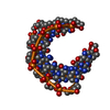
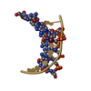
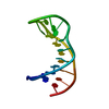
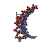
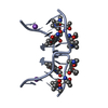
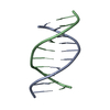
- PDB-481d: CRYSTAL STRUCTURE OF A HEXITOL NUCLEIC ACID (HNA) DUPLEX AT 1.6A ... -
+ Open data

- Basic information
| Entry | Database: PDB / ID: 481d | ||||||||||||||||||
|---|---|---|---|---|---|---|---|---|---|---|---|---|---|---|---|---|---|---|---|
| Title | CRYSTAL STRUCTURE OF A HEXITOL NUCLEIC ACID (HNA) DUPLEX AT 1.6A RESOLUTION | ||||||||||||||||||
 Components Components | 5'-H(* Keywords KeywordsDNA / HEXITOL NUCLEIC ACID / DNA ANALOGUE / DOUBLE HELIX | Function / homology | DNA |  Function and homology information Function and homology informationMethod |  X-RAY DIFFRACTION / SYNCHROTRON / Resolution: 1.6 Å X-RAY DIFFRACTION / SYNCHROTRON / Resolution: 1.6 Å  Authors AuthorsDeclercq, R. / Van Meervelt, L. |  CitationJournal: J.Am.Chem.Soc. / Year: 2002 CitationJournal: J.Am.Chem.Soc. / Year: 2002Title: Crystal structure of double helical hexitol nucleic acids. Authors: Declercq, R. / Van Aerschot, A. / Read, R.J. / Herdewijn, P. / Van Meervelt, L. #1: Journal: Angew.Chem.Int.Ed.Engl. / Year: 1995Title: 1',5'-Anhydrohexitol Nucleic Acids, a New Promising Antisense Construct Authors: Van Aerschot, A. / Verheggen, I. / Hendrix, C. / Herdewijn, P. #2: Journal: Acta Crystallogr.,Sect.C / Year: 1995Title: 1',5'-Anhydro-2',3'-Dideoxy-2'-(Guanin-9-Yl)-D-Arabino Hexitol Authors: Declercq, R. / Herdewijn, P. / Van Meervelt, L. #3: Journal: J.Am.Chem.Soc. / Year: 1998Title: Molecular Dynamics Simulation to Investigate Differences in Groove Hydration of HNA/RNA Hybrids as Compared to HNA/DNA Complexes Authors: De Winter, H. / Lescrinier, E. / Van Aerschot, A. / Herdewijn, P #4: Journal: Acta Crystallogr.,Sect.D / Year: 1999Title: Oligonucleotides with 1',5'-Anhydrohexitol Nucleoside Building Blocks: Crystallisation and Preliminary X-Ray Studies of h(GTGTACAC) Authors: Declercq, R. / Van Aerschot, A. / Herdewijn, P. / Van Meervelt, L. History |
|
- Structure visualization
| Structure viewer | Molecule: MolmilJmol/JSmol |
|---|
- Downloads & links
-Download
| PDBx/mmCIF format | 481d.cif.gz | 15.2 KB | Display | PDBx/mmCIF format |
|---|---|---|---|---|
| PDB format | pdb481d.ent.gz | 9.9 KB | Display | PDB format |
| PDBx/mmJSON format | 481d.json.gz | Tree view | PDBx/mmJSON format | |
| Others |  Other downloads Other downloads |
-Validation report
| Arichive directory | https://data.pdbj.org/pub/pdb/validation_reports/81/481dftp://data.pdbj.org/pub/pdb/validation_reports/81/481d | HTTPS FTP |
|---|
-Related structure data

-Links
 PDBj
PDBj

- Assembly
| Deposited unit | 
| ||||||||||
|---|---|---|---|---|---|---|---|---|---|---|---|
| 1 | 
| ||||||||||
| Unit cell |
|
-Components
| #1: DNA chain | Mass: 2538.831 Da / Num. of mol.: 1 / Source method: obtained synthetically Details: THE SUGAR MOIETIES ARE NOT DEOXYRIBOSE BUT D-ARABINO-HEXITOL |
|---|---|
| #2: Water | ChemComp-HOH /  Mass: 18.015 Da / Num. of mol.: 40 / Source method: isolated from a natural source / Formula: H2O Mass: 18.015 Da / Num. of mol.: 40 / Source method: isolated from a natural source / Formula: H2O |
-Experimental details
-Experiment
| Experiment | Method: X-RAY DIFFRACTION / Number of used crystals: 1 |
|---|
- Sample preparation
| Crystal | Density Matthews: 2.42 Å3/Da / Density % sol: 49.09 % | ||||||||||||||||||||||||||||||||||||||||||||||||||||||||
|---|---|---|---|---|---|---|---|---|---|---|---|---|---|---|---|---|---|---|---|---|---|---|---|---|---|---|---|---|---|---|---|---|---|---|---|---|---|---|---|---|---|---|---|---|---|---|---|---|---|---|---|---|---|---|---|---|---|
| Crystal grow | Method: vapor diffusion, hanging drop / pH: 7 Details: CRYSTALS WERE OBTAINED FROM A SOLUTION THAT CONTAINED CACODYLATE BUFFER, KCL, BACL2, SPERMINE TETRACHLORIDE, AND MPD, pH 7.0, VAPOR DIFFUSION, HANGING DROP | ||||||||||||||||||||||||||||||||||||||||||||||||||||||||
| Components of the solutions |
| ||||||||||||||||||||||||||||||||||||||||||||||||||||||||
| Crystal grow | *PLUS Temperature: 277 K | ||||||||||||||||||||||||||||||||||||||||||||||||||||||||
| Components of the solutions | *PLUS
|
-Data collection
| Diffraction | Mean temperature: 100 K |
|---|---|
| Diffraction source | Source: SYNCHROTRON / Site: EMBL/DESY, HAMBURG  / Beamline: BW7B / Wavelength: 0.8375 / Beamline: BW7B / Wavelength: 0.8375 |
| Detector | Type: MARRESEARCH / Detector: IMAGE PLATE / Date: Oct 22, 1998 |
| Radiation | Monochromator: MIRRORS / Protocol: SINGLE WAVELENGTH / Monochromatic (M) / Laue (L): M / Scattering type: x-ray |
| Radiation wavelength | Wavelength: 0.8375 Å / Relative weight: 1 |
| Reflection | Resolution: 1.6→20 Å / Num. all: 3345 / Num. obs: 3345 / % possible obs: 99.9 % / Observed criterion σ(I): 0 / Redundancy: 8.5 % / Rmerge(I) obs: 0.071 / Net I/σ(I): 13.2 |
| Reflection shell | Resolution: 1.6→1.63 Å / Redundancy: 4.4 % / Rmerge(I) obs: 0.173 / Mean I/σ(I) obs: 6.6 / % possible all: 100 |
| Reflection | *PLUS Lowest resolution: 20 Å / Rmerge(I) obs: 0.071 |
| Reflection shell | *PLUS % possible obs: 100 % / Rmerge(I) obs: 0.173 |
- Processing
| Software |
| |||||||||||||||||||||||||||||||||
|---|---|---|---|---|---|---|---|---|---|---|---|---|---|---|---|---|---|---|---|---|---|---|---|---|---|---|---|---|---|---|---|---|---|---|
| Refinement | Starting model: 2.21A P 62 2 2 STRUCTURE OF H(GTGTACAC) (NDB CODE HD0002) WAS USED AS MODEL Resolution: 1.6→20 Å / Num. parameters: 843 / Num. restraintsaints: 1426 / Cross valid method: FREE R / σ(F): 0 StereochEM target val spec case: ALL OTHER CHEMICALLY EQUIVALENT BOND DISTANCES AND ANGLES WERE RESTRAINED TO BE SIMILAR BY SAME DISTANCE RESTRAINTS
| |||||||||||||||||||||||||||||||||
| Solvent computation | Solvent model: BULK SOLVENT MODELING: MOEWS & KRETSINGER, J.MOL.BIOL. 91, 201-228 (1973) | |||||||||||||||||||||||||||||||||
| Refine analyze | Occupancy sum hydrogen: 0 / Occupancy sum non hydrogen: 210 | |||||||||||||||||||||||||||||||||
| Refinement step | Cycle: LAST / Resolution: 1.6→20 Å
| |||||||||||||||||||||||||||||||||
| Refine LS restraints |
| |||||||||||||||||||||||||||||||||
| Software | *PLUS Name: SHELXL / Version: 97 / Classification: refinement | |||||||||||||||||||||||||||||||||
| Refinement | *PLUS Rfactor all: 0.1835 / Rfactor obs: 0.174 / Rfactor Rfree: 0.229 / Rfactor Rwork: 0.174 | |||||||||||||||||||||||||||||||||
| Solvent computation | *PLUS | |||||||||||||||||||||||||||||||||
| Displacement parameters | *PLUS |
