 Movie
Movie Controller

[English] 日本語
 Yorodumi
- PDB-1d7z: CRYSTAL STRUCTURE OF A HEXITOL NUCLEIC ACID (HNA) DUPLEX AT 2.2 A... -
+ Open data

- Basic information
| Entry | Database: PDB / ID: 1d7z | ||||||||||||||||||
|---|---|---|---|---|---|---|---|---|---|---|---|---|---|---|---|---|---|---|---|
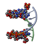
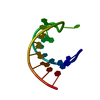
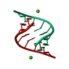
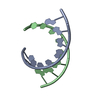
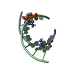
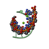
| Title | CRYSTAL STRUCTURE OF A HEXITOL NUCLEIC ACID (HNA) DUPLEX AT 2.2 A RESOLUTION | ||||||||||||||||||
 Components Components | 5'-H(* Keywords KeywordsDNA / HEXITOL NUCLEIC ACID / DNA ANALOGUE / DOUBLE HELIX | Function / homology | DNA |  Function and homology information Function and homology informationMethod |  X-RAY DIFFRACTION / SYNCHROTRON / Resolution: 2.21 Å X-RAY DIFFRACTION / SYNCHROTRON / Resolution: 2.21 Å  Authors AuthorsDeclercq, R. / Van Meervelt, L. |  CitationJournal: J.Am.Chem.Soc. / Year: 2002 CitationJournal: J.Am.Chem.Soc. / Year: 2002Title: Crystal structure of double helical hexitol nucleic acids. Authors: Declercq, R. / Van Aerschot, A. / Read, R.J. / Herdewijn, P. / Van Meervelt, L. #1: Journal: Angew.Chem.Int.Ed.Engl. / Year: 1995Title: 1',5'-Anhydrohexitol nucleic acids, a new promising antisense construct Authors: Van Aerschot, A. / Verheggen, I. / Hendrix, C. / Herdewijn, P. #2: Journal: Acta Crystallogr.,Sect.D / Year: 1995Title: 1',5'-anhydro-2',3'-dideoxy-2'-(guanin-9-yl)-D-arabino-hexitol Authors: Declercq, R. / Herdewijn, P. / Van Meervelt, L. #3: Journal: Acta Crystallogr.,Sect.D / Year: 1999Title: Oligonucleotides with 1',5'-anhydrohexitol nucleoside building blocks: Crystallization and preliminary X-ray studies of H(GTGTACAC) Authors: Declercq, R. / Van Aerschot, A. / Herdewijn, P. / Van Meervelt, L. History |
|
- Structure visualization
| Structure viewer | Molecule: MolmilJmol/JSmol |
|---|
- Downloads & links
-Download
| PDBx/mmCIF format | 1d7z.cif.gz | 15 KB | Display | PDBx/mmCIF format |
|---|---|---|---|---|
| PDB format | pdb1d7z.ent.gz | 10.1 KB | Display | PDB format |
| PDBx/mmJSON format | 1d7z.json.gz | Tree view | PDBx/mmJSON format | |
| Others |  Other downloads Other downloads |
-Validation report
| Arichive directory | https://data.pdbj.org/pub/pdb/validation_reports/d7/1d7zftp://data.pdbj.org/pub/pdb/validation_reports/d7/1d7z | HTTPS FTP |
|---|
-Related structure data
| Related structure data |  481dC C: citing same article ( |
|---|---|
| Similar structure data | |
| Other databases |
|


-Links
 PDBj
PDBj

- Assembly
| Deposited unit | 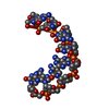
| ||||||||||
|---|---|---|---|---|---|---|---|---|---|---|---|
| 1 | 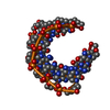
| ||||||||||
| Unit cell |
| ||||||||||
| Components on special symmetry positions |
|
-Components
| #1: DNA chain | Mass: 2538.831 Da / Num. of mol.: 1 / Source method: obtained synthetically Details: THE MOLECULE WAS CHEMICALLY SYNTESISED (SEE REFERENCE 1) |
|---|---|
| #2: Water | ChemComp-HOH /  Mass: 18.015 Da / Num. of mol.: 38 / Source method: isolated from a natural source / Formula: H2O Mass: 18.015 Da / Num. of mol.: 38 / Source method: isolated from a natural source / Formula: H2O |
-Experimental details
-Experiment
| Experiment | Method: X-RAY DIFFRACTION / Number of used crystals: 1 |
|---|
- Sample preparation
| Crystal | Density Matthews: 2.6 Å3/Da / Density % sol: 52.66 % | ||||||||||||||||||||||||||||||||||||||||||||||||||||||||
|---|---|---|---|---|---|---|---|---|---|---|---|---|---|---|---|---|---|---|---|---|---|---|---|---|---|---|---|---|---|---|---|---|---|---|---|---|---|---|---|---|---|---|---|---|---|---|---|---|---|---|---|---|---|---|---|---|---|
| Crystal grow | Temperature: 277 K / Method: vapor diffusion, hanging drop / pH: 7 Details: CACODYLATE BUFFER, KCL, BACL2, SPERMINE TETRACHLORIDE AND MPD, pH 7.0, VAPOR DIFFUSION, HANGING DROP at 277K | ||||||||||||||||||||||||||||||||||||||||||||||||||||||||
| Components of the solutions |
| ||||||||||||||||||||||||||||||||||||||||||||||||||||||||
| Crystal grow | *PLUS Temperature: 277 K / Method: vapor diffusion, hanging drop | ||||||||||||||||||||||||||||||||||||||||||||||||||||||||
| Components of the solutions | *PLUS
|
-Data collection
| Diffraction | Mean temperature: 100 K |
|---|---|
| Diffraction source | Source: SYNCHROTRON / Site: EMBL/DESY, HAMBURG  / Beamline: X11 / Wavelength: 0.9116 / Beamline: X11 / Wavelength: 0.9116 |
| Detector | Type: MARRESEARCH / Detector: IMAGE PLATE / Date: Apr 11, 1999 |
| Radiation | Protocol: SINGLE WAVELENGTH / Monochromatic (M) / Laue (L): M / Scattering type: x-ray |
| Radiation wavelength | Wavelength: 0.9116 Å / Relative weight: 1 |
| Reflection | Resolution: 2.21→20 Å / Num. all: 1344 / Num. obs: 1344 / % possible obs: 83.8 % / Observed criterion σ(I): 0 / Redundancy: 16.8 % / Biso Wilson estimate: 50.8 Å2 / Rmerge(I) obs: 0.051 / Net I/σ(I): 38.8 |
| Reflection shell | Resolution: 2.21→2.25 Å / Rmerge(I) obs: 0.168 / % possible all: 74.1 |
| Reflection | *PLUS Rmerge(I) obs: 0.051 |
| Reflection shell | *PLUS % possible obs: 74.1 % / Rmerge(I) obs: 0.168 |
- Processing
| Software |
| |||||||||||||||||||||||||||||||||
|---|---|---|---|---|---|---|---|---|---|---|---|---|---|---|---|---|---|---|---|---|---|---|---|---|---|---|---|---|---|---|---|---|---|---|
| Refinement | Resolution: 2.21→20 Å / Cross valid method: NONE USED / σ(F): 0 / σ(I): 0 Stereochemistry target values: BASES: TAYLOR AND KENNARD VALUES; THE REST: VALUES BASED ON SINGLE CRYSTAL STRUCTURE OF HNA BUILDING BLOCKS
| |||||||||||||||||||||||||||||||||
| Refinement step | Cycle: LAST / Resolution: 2.21→20 Å
| |||||||||||||||||||||||||||||||||
| Refine LS restraints |
| |||||||||||||||||||||||||||||||||
| Software | *PLUS Name: SHELXL / Version: 97 / Classification: refinement | |||||||||||||||||||||||||||||||||
| Refinement | *PLUS Lowest resolution: 10 Å / Rfactor all: 0.233 / Rfactor obs: 0.233 / Rfactor Rwork: 0.232 | |||||||||||||||||||||||||||||||||
| Solvent computation | *PLUS | |||||||||||||||||||||||||||||||||
| Displacement parameters | *PLUS |
