 Movie
Movie Controller

[English] 日本語
 Yorodumi
- PDB-3zzz: Crystal structure of a Raver1 PRI4 peptide in complex with polypy... -
+ Open data

- Basic information
| Entry | Database: PDB / ID: 3zzz | ||||||
|---|---|---|---|---|---|---|---|
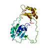
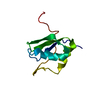
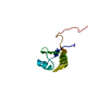
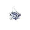
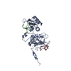
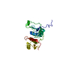
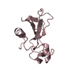
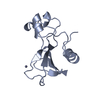
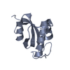
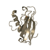
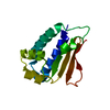
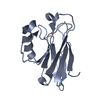
| Title | Crystal structure of a Raver1 PRI4 peptide in complex with polypyrimidine tract binding protein RRM2 | ||||||
 Components Components |
| ||||||
 Keywords Keywords | PROTEIN BINDING / PEPTIDE BINDING / RNA RECOGNITION MOTIF | ||||||
| Function / homology |  Function and homology information Function and homology informationnegative regulation of muscle cell differentiation / poly-pyrimidine tract binding / IRES-dependent viral translational initiation / positive regulation of calcineurin-NFAT signaling cascade / pre-mRNA binding / FGFR2 alternative splicing / negative regulation of RNA splicing / negative regulation of neuron differentiation / regulation of alternative mRNA splicing, via spliceosome / regulation of RNA splicing ...negative regulation of muscle cell differentiation / poly-pyrimidine tract binding / IRES-dependent viral translational initiation / positive regulation of calcineurin-NFAT signaling cascade / pre-mRNA binding / FGFR2 alternative splicing / negative regulation of RNA splicing / negative regulation of neuron differentiation / regulation of alternative mRNA splicing, via spliceosome / regulation of RNA splicing / negative regulation of mRNA splicing, via spliceosome / Processing of Capped Intron-Containing Pre-mRNA / regulation of cell differentiation / mRNA Splicing - Major Pathway / neurogenesis / RNA splicing / mRNA processing / mRNA binding / nucleolus / positive regulation of transcription by RNA polymerase II / RNA binding / extracellular exosome / nucleoplasm / membrane / nucleus / cytoplasm Similarity search - Function | ||||||
| Biological species |  HOMO SAPIENS (human) HOMO SAPIENS (human) | ||||||
| Method |  X-RAY DIFFRACTION / SYNCHROTRON / MOLECULAR REPLACEMENT / Resolution: 1.55 Å X-RAY DIFFRACTION / SYNCHROTRON / MOLECULAR REPLACEMENT / Resolution: 1.55 Å | ||||||
 Authors Authors | Joshi, A. / Kotik-Kogan, O. / Curry, S. | ||||||
 Citation Citation | Journal: Structure / Year: 2011 Title: Crystallographic Analysis of Polypyrimidine Tract-Binding Protein-Raver1 Interactions Involved in Regulation of Alternative Splicing. Authors: Joshi, A. / Coelho, M.B. / Kotik-Kogan, O. / Simpson, P.J. / Matthews, S.J. / Smith, C.W. / Curry, S. | ||||||
| History |
|
- Structure visualization
| Structure viewer | Molecule: MolmilJmol/JSmol |
|---|
- Downloads & links
-Download
| PDBx/mmCIF format | 3zzz.cif.gz | 60.2 KB | Display | PDBx/mmCIF format |
|---|---|---|---|---|
| PDB format | pdb3zzz.ent.gz | 43.2 KB | Display | PDB format |
| PDBx/mmJSON format | 3zzz.json.gz | Tree view | PDBx/mmJSON format | |
| Others |  Other downloads Other downloads |
-Validation report
| Arichive directory | https://data.pdbj.org/pub/pdb/validation_reports/zz/3zzzftp://data.pdbj.org/pub/pdb/validation_reports/zz/3zzz | HTTPS FTP |
|---|
-Related structure data
| Related structure data |  3zzySC S: Starting model for refinement C: citing same article ( |
|---|---|
| Similar structure data |


-Links
 PDBj
PDBj

- Assembly
| Deposited unit | 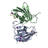
| ||||||||
|---|---|---|---|---|---|---|---|---|---|
| 1 | 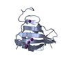
| ||||||||
| 2 | 
| ||||||||
| Unit cell |
| ||||||||
| Noncrystallographic symmetry (NCS) | NCS oper: (Code: given Matrix: (-0.1983, 0.03711, 0.9794), Vector: |
-Components
| #1: Protein | Mass: 14169.072 Da / Num. of mol.: 2 / Fragment: RNA RECOGNITION MOTIF 2, RESIDUES 156-285 Source method: isolated from a genetically manipulated source Source: (gene. exp.) HOMO SAPIENS (human) / Production host:  #2: Protein/peptide | Mass: 1399.572 Da / Num. of mol.: 2 / Fragment: MOTIF PRI4, RESIDUES 680-692 Source method: isolated from a genetically manipulated source Details: THIS FRAGMENT IS ACTUALLY FUSED TO THE N-TERMINUS OF MOLECULE 1, SEE REMARK 999 Source: (gene. exp.) #3: Chemical | ChemComp-IOD /   Mass: 126.904 Da / Num. of mol.: 5 / Source method: obtained synthetically / Formula: I Mass: 126.904 Da / Num. of mol.: 5 / Source method: obtained synthetically / Formula: I#4: Water | ChemComp-HOH / |  Mass: 18.015 Da / Num. of mol.: 132 / Source method: isolated from a natural source / Formula: H2O Mass: 18.015 Da / Num. of mol.: 132 / Source method: isolated from a natural source / Formula: H2OHas protein modification | Y | Sequence details | FIRST 3 RESIDUES (GAM) ARE VECTOR DERIVED. RESIDUES 4 - 16 CORRESPOND TO RESIDUES 680-692 OF RAVER1 ...FIRST 3 RESIDUES (GAM) ARE VECTOR DERIVED. RESIDUES 4 - 16 CORRESPOND | |
|---|
-Experimental details
-Experiment
| Experiment | Method: X-RAY DIFFRACTION / Number of used crystals: 1 |
|---|
- Sample preparation
| Crystal | Density Matthews: 2.01 Å3/Da / Density % sol: 38.94 % / Description: NONE |
|---|---|
| Crystal grow | pH: 6.5 / Details: SEE PAPER., pH 6.5 |
-Data collection
| Diffraction | Mean temperature: 100 K |
|---|---|
| Diffraction source | Source: SYNCHROTRON / Site: Diamond  / Beamline: I02 / Wavelength: 0.9796 / Beamline: I02 / Wavelength: 0.9796 |
| Detector | Type: ADSC QUANTUM 315r / Detector: CCD / Date: Sep 24, 2009 / Details: MIRRORS |
| Radiation | Monochromator: DOUBLE CRYSTAL MONOCHROMATOR / Protocol: SINGLE WAVELENGTH / Monochromatic (M) / Laue (L): M / Scattering type: x-ray |
| Radiation wavelength | Wavelength: 0.9796 Å / Relative weight: 1 |
| Reflection | Resolution: 1.55→35.49 Å / Num. obs: 36184 / % possible obs: 96.9 % / Observed criterion σ(I): 0 / Redundancy: 2.4 % / Biso Wilson estimate: 21.5 Å2 / Rmerge(I) obs: 0.06 / Net I/σ(I): 10.2 |
| Reflection shell | Resolution: 1.55→1.63 Å / Redundancy: 2.4 % / Rmerge(I) obs: 0.36 / Mean I/σ(I) obs: 3 / % possible all: 96.5 |
- Processing
| Software |
| ||||||||||||||||||||||||||||||||||||||||||||||||||||||||||||
|---|---|---|---|---|---|---|---|---|---|---|---|---|---|---|---|---|---|---|---|---|---|---|---|---|---|---|---|---|---|---|---|---|---|---|---|---|---|---|---|---|---|---|---|---|---|---|---|---|---|---|---|---|---|---|---|---|---|---|---|---|---|
| Refinement | Method to determine structure: MOLECULAR REPLACEMENT Starting model: PDB ENTRY 3ZZY Resolution: 1.55→20.07 Å / Rfactor Rfree error: 0.005 / Data cutoff high absF: 1123728.72 / Data cutoff low absF: 0 / Cross valid method: THROUGHOUT / σ(F): 0 / Stereochemistry target values: MLF Details: BULK SOLVENT MODEL USED. THE CRYSTALLISED PROTEIN HAS A RAVER1 PEPTIDE GENETICALLY FUSED AS AN N-TERMINAL EXTENSION OF PTB RRM3. OWING TO LINKER DISORDER WE CANNOT DETERMINE WHICH RRM DOMAIN ...Details: BULK SOLVENT MODEL USED. THE CRYSTALLISED PROTEIN HAS A RAVER1 PEPTIDE GENETICALLY FUSED AS AN N-TERMINAL EXTENSION OF PTB RRM3. OWING TO LINKER DISORDER WE CANNOT DETERMINE WHICH RRM DOMAIN (CHAINS A, B) IS COVALENTLY LINKED TO WHICH RAVER1 PEPTIDE (CHAINS C,D).
| ||||||||||||||||||||||||||||||||||||||||||||||||||||||||||||
| Solvent computation | Solvent model: FLAT MODEL / Bsol: 67.8846 Å2 / ksol: 0.5 e/Å3 | ||||||||||||||||||||||||||||||||||||||||||||||||||||||||||||
| Displacement parameters | Biso mean: 21.6 Å2
| ||||||||||||||||||||||||||||||||||||||||||||||||||||||||||||
| Refine analyze |
| ||||||||||||||||||||||||||||||||||||||||||||||||||||||||||||
| Refinement step | Cycle: LAST / Resolution: 1.55→20.07 Å
| ||||||||||||||||||||||||||||||||||||||||||||||||||||||||||||
| Refine LS restraints |
| ||||||||||||||||||||||||||||||||||||||||||||||||||||||||||||
| LS refinement shell | Resolution: 1.55→1.65 Å / Rfactor Rfree error: 0.02 / Total num. of bins used: 6
| ||||||||||||||||||||||||||||||||||||||||||||||||||||||||||||
| Xplor file |
|
