- PDB-3zx7: Complex of lysenin with phosphocholine -
+
Open data
ID or keywords:
Loading...
-
Basic information
Entry
Database: PDB / ID: 3zx7
Title
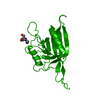
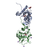
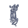
Complex of lysenin with phosphocholine
Components
LYSENIN
Keywords
TOXIN / PORE FORMING TOXIN
Function / homology
Function and homology information
other organism cell membrane / monoatomic ion transport / toxin activity / killing of cells of another organism / defense response to bacterium / extracellular region Similarity search - Function
Resolution: 2.8→32.1 Å / Num. obs: 12994 / % possible obs: 99.9 % / Observed criterion σ(I): 6 / Redundancy: 22.1 % / Biso Wilson estimate: 72.61 Å2 / Rmerge(I) obs: 0.1 / Net I/σ(I): 29
Reflection shell
Resolution: 2.8→2.9 Å / Redundancy: 23 % / Rmerge(I) obs: 0.8 / Mean I/σ(I) obs: 4.9 / % possible all: 99.9
-
Processing
Software
Name
Version
Classification
BUSTER
2.11.1
refinement
XDS
datareduction
SCALA
datascaling
SHARP
phasing
Refinement
Method to determine structure: MIRAS Starting model: NONE Resolution: 2.84→23.7 Å / Cor.coef. Fo:Fc: 0.8919 / Cor.coef. Fo:Fc free: 0.8532 / Cross valid method: THROUGHOUT / σ(F): 0 / SU R Blow DPI: 0.628 / SU Rfree Blow DPI: 0.33 Details: IDEAL-DIST CONTACT TERM CONTACT SETUP. RESIDUE TYPES WITHOUT CCP4 ATOM TYPE IN LIBRARY=PO4 PC. NUMBER OF ATOMS WITH PROPER CCP4 ATOM TYPE=4713. NUMBER WITH APPROX DEFAULT CCP4 ATOM TYPE=58. ...Details: IDEAL-DIST CONTACT TERM CONTACT SETUP. RESIDUE TYPES WITHOUT CCP4 ATOM TYPE IN LIBRARY=PO4 PC. NUMBER OF ATOMS WITH PROPER CCP4 ATOM TYPE=4713. NUMBER WITH APPROX DEFAULT CCP4 ATOM TYPE=58. NUMBER TREATED BY BAD NON-BONDED CONTACTS=6.
Rfactor
Num. reflection
% reflection
Selection details
Rfree
0.264
630
4.87 %
RANDOM
Rwork
0.2235
-
-
-
obs
0.2255
12930
99.57 %
-
Displacement parameters
Biso mean: 75.88 Å2
Baniso -1
Baniso -2
Baniso -3
1-
-3.3721 Å2
0 Å2
0 Å2
2-
-
-3.3721 Å2
0 Å2
3-
-
-
6.7443 Å2
Refine analyze
Luzzati coordinate error obs: 0.499 Å
Refinement step
Cycle: LAST / Resolution: 2.84→23.7 Å
Protein
Nucleic acid
Ligand
Solvent
Total
Num. atoms
2355
0
72
24
2451
Refine LS restraints
Refine-ID
Type
Dev ideal
Number
Restraint function
Weight
X-RAY DIFFRACTION
t_bond_d
0.009
4770
HARMONIC
2
X-RAY DIFFRACTION
t_angle_deg
1.11
8576
HARMONIC
2
X-RAY DIFFRACTION
t_dihedral_angle_d
1037
SINUSOIDAL
2
X-RAY DIFFRACTION
t_incorr_chiral_ct
X-RAY DIFFRACTION
t_pseud_angle
X-RAY DIFFRACTION
t_trig_c_planes
57
HARMONIC
2
X-RAY DIFFRACTION
t_gen_planes
698
HARMONIC
5
X-RAY DIFFRACTION
t_it
4770
HARMONIC
20
X-RAY DIFFRACTION
t_nbd
0
SEMIHARMONIC
5
X-RAY DIFFRACTION
t_omega_torsion
2.97
X-RAY DIFFRACTION
t_other_torsion
18.06
X-RAY DIFFRACTION
t_improper_torsion
X-RAY DIFFRACTION
t_chiral_improper_torsion
323
SEMIHARMONIC
5
X-RAY DIFFRACTION
t_sum_occupancies
X-RAY DIFFRACTION
t_utility_distance
X-RAY DIFFRACTION
t_utility_angle
X-RAY DIFFRACTION
t_utility_torsion
X-RAY DIFFRACTION
t_ideal_dist_contact
5048
SEMIHARMONIC
4
LS refinement shell
Resolution: 2.84→3.07 Å / Total num. of bins used: 7
Rfactor
Num. reflection
% reflection
Rfree
0.3229
133
5.18 %
Rwork
0.2286
2435
-
all
0.2335
2568
-
obs
-
-
99.57 %
Refinement TLS params.
Method: refined / Refine-ID: X-RAY DIFFRACTION
ID
L11 (°2)
L12 (°2)
L13 (°2)
L22 (°2)
L23 (°2)
L33 (°2)
S11 (Å °)
S12 (Å °)
S13 (Å °)
S21 (Å °)
S22 (Å °)
S23 (Å °)
S31 (Å °)
S32 (Å °)
S33 (Å °)
T11 (Å2)
T12 (Å2)
T13 (Å2)
T22 (Å2)
T23 (Å2)
T33 (Å2)
Origin x (Å)
Origin y (Å)
Origin z (Å)
1
12.8321
2.4157
-0.1397
1.2612
-2.0132
0
-0.211
0.0907
-0.2162
0.1033
0.3763
-1.6083
0.005
0.6495
-0.1653
-0.133
0.0349
-0.1579
-0.4334
-0.2936
0.3484
36.9913
10.9084
15.2861
2
-0.1155
0.4914
0.3796
0.6059
-1.5738
0
-0.2287
0.2949
0.0611
-0.0399
0.101
-0.5392
0.2048
-0.1144
0.1277
-0.3839
-0.0919
0.0744
-0.0269
-0.4529
0.6352
58.6132
21.7952
4.3616
3
6.7762
3.9792
2.2074
8.2472
6.8256
9.0139
-0.1156
0.9431
-0.3629
-0.0787
0.1539
-1.2146
-0.0118
-0.5225
-0.0383
-0.4242
0.1324
-0.1354
-0.2475
-0.1105
-0.0009
30.7287
13.9571
10.881
4
6.8949
-0.0401
-1.0641
0.376
1.7531
2.9648
-0.2212
0.2427
-0.3622
-0.261
-0.2602
-0.024
0.198
0.1932
0.4815
-0.2545
0.227
-0.0545
-0.2229
-0.296
0.6399
52.5122
16.3965
12.8242
5
-2.76
0.743
-1.9525
13.4924
-5.3695
1.4581
0.0384
0.0624
-0.3023
-0.1359
0.3202
0.3283
-0.1374
0.1293
-0.3586
-0.1918
0.4502
0.2971
0.3413
0.1111
0.5471
63.7172
9.8669
10.4798
6
-2.9285
3.2837
3.2147
6.6883
6.3271
0.3673
0.0566
0.0516
-0.0405
-0.1242
-0.1538
-0.0281
0.249
-0.0337
0.0973
0.3011
0.1539
-0.4426
-0.1409
-0.1453
0.7294
42.452
6.4808
19.0233
7
1.752
-0.1445
1.5569
2.7107
0.1655
5.6395
-0.0354
-0.1959
0.3384
0.4458
-0.3301
0.0561
-0.2518
-0.6024
0.3655
0.0835
0.0443
-0.039
-0.1338
-0.1257
-0.2682
5.2741
14.3894
9.0919
8
2.6507
1.9963
2.4964
6.8664
0.5138
5.9942
-0.2072
0.1756
0.7188
-0.0906
-0.1854
-0.2316
-1.2995
-0.3317
0.3926
0.0875
0.1041
-0.1652
-0.1687
-0.0214
-0.2662
4.3686
22.3767
-2.2726
9
2.239
-1.9796
0.4614
0.1405
0.0837
0.1119
-0.167
0.0906
0.2998
0.2492
-0.1408
-0.0099
0.1744
-0.2178
0.3078
0.2976
0.0114
-0.1044
-0.169
-0.0426
-0.1189
11.8308
17.9678
13.1992
Refinement TLS group
ID
Refine-ID
Refine TLS-ID
Selection details
1
X-RAY DIFFRACTION
1
CHAINAANDRESID6:22
2
X-RAY DIFFRACTION
2
CHAINAANDRESID23:48
3
X-RAY DIFFRACTION
3
CHAINAANDRESID49:75
4
X-RAY DIFFRACTION
4
CHAINAANDRESID76:131
5
X-RAY DIFFRACTION
5
CHAINAANDRESID132:147
6
X-RAY DIFFRACTION
6
CHAINAANDRESID148:154
7
X-RAY DIFFRACTION
7
CHAINAANDRESID155:267
8
X-RAY DIFFRACTION
8
CHAINAANDRESID268:286
9
X-RAY DIFFRACTION
9
CHAINAANDRESID287:992
+
About Yorodumi
-
News
-
Feb 9, 2022. New format data for meta-information of EMDB entries
New format data for meta-information of EMDB entries
Version 3 of the EMDB header file is now the official format.
The previous official version 1.9 will be removed from the archive.
In the structure databanks used in Yorodumi, some data are registered as the other names, "COVID-19 virus" and "2019-nCoV". Here are the details of the virus and the list of structure data.
Jan 31, 2019. EMDB accession codes are about to change! (news from PDBe EMDB page)
EMDB accession codes are about to change! (news from PDBe EMDB page)
The allocation of 4 digits for EMDB accession codes will soon come to an end. Whilst these codes will remain in use, new EMDB accession codes will include an additional digit and will expand incrementally as the available range of codes is exhausted. The current 4-digit format prefixed with “EMD-” (i.e. EMD-XXXX) will advance to a 5-digit format (i.e. EMD-XXXXX), and so on. It is currently estimated that the 4-digit codes will be depleted around Spring 2019, at which point the 5-digit format will come into force.
The EM Navigator/Yorodumi systems omit the EMD- prefix.
Related info.:Q: What is EMD? / ID/Accession-code notation in Yorodumi/EM Navigator
Yorodumi is a browser for structure data from EMDB, PDB, SASBDB, etc.
This page is also the successor to EM Navigator detail page, and also detail information page/front-end page for Omokage search.
The word "yorodu" (or yorozu) is an old Japanese word meaning "ten thousand". "mi" (miru) is to see.
Related info.:EMDB / PDB / SASBDB / Comparison of 3 databanks / Yorodumi Search / Aug 31, 2016. New EM Navigator & Yorodumi / Yorodumi Papers / Jmol/JSmol / Function and homology information / Changes in new EM Navigator and Yorodumi
 Movie
Movie Controller


 Open data
 Basic information
 Components
 Keywords
 Function and homology information
 X-RAY DIFFRACTION /
  Authors
 Citation
 Structure visualization
 Downloads & links
 Other downloads
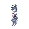


 PDBj
PDBj Assembly


 Mass: 94.971 Da / Num. of mol.: 5 / Source method: obtained synthetically / Formula: PO4
 Mass: 184.151 Da / Num. of mol.: 4 / Source method: obtained synthetically / Formula: C5H15NO4P
 Mass: 22.990 Da / Num. of mol.: 3 / Source method: obtained synthetically / Formula: Na
 Mass: 18.015 Da / Num. of mol.: 24 / Source method: isolated from a natural source / Formula: H2O
 Sample preparation
 / Beamline: BM14 / Wavelength: 0.97867
  Processing