 Movie
Movie Controller

[English] 日本語
 Yorodumi
- PDB-3zdr: Structure of the Alcohol dehydrogenase (ADH) domain of a bifuncti... -
+ Open data

- Basic information
| Entry | Database: PDB / ID: 3zdr | |||||||||
|---|---|---|---|---|---|---|---|---|---|---|
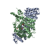
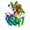
| Title | Structure of the Alcohol dehydrogenase (ADH) domain of a bifunctional ADHE dehydrogenase from Geobacillus thermoglucosidasius NCIMB 11955 | |||||||||
 Components Components | ALCOHOL DEHYDROGENASE DOMAIN OF THE BIFUNCTIONAL ACETALDEHYDE DEHYDROGENASE | |||||||||
 Keywords Keywords | OXIDOREDUCTASE / BIFUNCTIONAL ALCOHOL/ALDEHYDE DEHYDROGENASE / BIOETHANOL | |||||||||
| Function / homology |  Function and homology information Function and homology informationacetaldehyde dehydrogenase (acetylating) activity / alcohol metabolic process / carbon utilization / alcohol dehydrogenase (NAD+) activity / metal ion binding Similarity search - Function | |||||||||
| Biological species |  GEOBACILLUS THERMOGLUCOSIDASIUS (bacteria) GEOBACILLUS THERMOGLUCOSIDASIUS (bacteria) | |||||||||
| Method |  X-RAY DIFFRACTION / SYNCHROTRON / MOLECULAR REPLACEMENT / Resolution: 2.504 Å X-RAY DIFFRACTION / SYNCHROTRON / MOLECULAR REPLACEMENT / Resolution: 2.504 Å | |||||||||
 Authors Authors | Extance, J. / Crennell, S.J. / Eley, K. / Cripps, R. / Hough, D.W. / Danson, M.J. | |||||||||
 Citation Citation | Journal: Acta Crystallogr.,Sect.D / Year: 2013 Title: Structure of a Bifunctional Alcohol Dehydrogenase Involved in Bioethanol Generation in Geobacillus Thermoglucosidasius Authors: Extance, J. / Crennell, S.J. / Eley, K. / Cripps, R. / Hough, D.W. / Danson, M.J. | |||||||||
| History |
|
- Structure visualization
| Structure viewer | Molecule: MolmilJmol/JSmol |
|---|
- Downloads & links
-Download
| PDBx/mmCIF format | 3zdr.cif.gz | 161.8 KB | Display | PDBx/mmCIF format |
|---|---|---|---|---|
| PDB format | pdb3zdr.ent.gz | 130 KB | Display | PDB format |
| PDBx/mmJSON format | 3zdr.json.gz | Tree view | PDBx/mmJSON format | |
| Others |  Other downloads Other downloads |
-Validation report
| Arichive directory | https://data.pdbj.org/pub/pdb/validation_reports/zd/3zdrftp://data.pdbj.org/pub/pdb/validation_reports/zd/3zdr | HTTPS FTP |
|---|
-Related structure data
| Related structure data |  1rrmS S: Starting model for refinement |
|---|---|
| Similar structure data |


-Links
 PDBj
PDBj

- Assembly
| Deposited unit | 
| ||||||||
|---|---|---|---|---|---|---|---|---|---|
| 1 | 
| ||||||||
| Unit cell |
|
-Components
| #1: Protein | Mass: 48693.152 Da / Num. of mol.: 1 / Fragment: RESIDUES 457-867 Source method: isolated from a genetically manipulated source Details: C-TERMINAL ADH DOMAIN, STARTING AT RESIDUE 459 Source: (gene. exp.) GEOBACILLUS THERMOGLUCOSIDASIUS (bacteria)Strain: TM242 Description: THE NCIMB 11955 STRAIN ENGINEERED BY TMO RENEWABLES LTD TO INCREASE ETHANOL PRODUCTION. Production host: References: UniProt: F8CW95, acetaldehyde dehydrogenase (acetylating), alcohol dehydrogenase |
|---|---|
| #2: Chemical | ChemComp-ZN /   Mass: 65.409 Da / Num. of mol.: 1 / Source method: obtained synthetically / Formula: Zn Mass: 65.409 Da / Num. of mol.: 1 / Source method: obtained synthetically / Formula: Zn |
| #3: Chemical | ChemComp-SO4 /   Mass: 96.063 Da / Num. of mol.: 1 / Source method: obtained synthetically / Formula: SO4 Mass: 96.063 Da / Num. of mol.: 1 / Source method: obtained synthetically / Formula: SO4 |
| #4: Chemical | ChemComp-GOL /   Mass: 92.094 Da / Num. of mol.: 1 / Source method: obtained synthetically / Formula: C3H8O3 Mass: 92.094 Da / Num. of mol.: 1 / Source method: obtained synthetically / Formula: C3H8O3 |
| #5: Water | ChemComp-HOH /  Mass: 18.015 Da / Num. of mol.: 27 / Source method: isolated from a natural source / Formula: H2O Mass: 18.015 Da / Num. of mol.: 27 / Source method: isolated from a natural source / Formula: H2O |
| Has protein modification | Y |
| Sequence details | NON-PUBLISHED GENOME. THE PROTEIN COMES FROM A DIFFERENT STRAIN OF GEOBACILLU |
-Experimental details
-Experiment
| Experiment | Method: X-RAY DIFFRACTION / Number of used crystals: 2 |
|---|
- Sample preparation
| Crystal | Density Matthews: 2.1 Å3/Da / Density % sol: 42 % / Description: NONE |
|---|---|
| Crystal grow | pH: 5 Details: 0.1 M SODIUM ACETATE PH 5.0, 0.1 M AMMONIUM SULPHATE, 0.3 M SODIUM FORMATE, 11.5% (V/V) PEG2K MME, AND 3-3.5% (V/V) PGA-LM (30% GLYCEROL CRYOPROTECTANT) |
-Data collection
| Diffraction | Mean temperature: 100 K |
|---|---|
| Diffraction source | Source: SYNCHROTRON / Site: Diamond  / Beamline: I03 / Wavelength: 0.97625 / Beamline: I03 / Wavelength: 0.97625 |
| Detector | Type: ADSC QUANTUM 315 / Detector: CCD / Date: Jan 30, 2011 / Details: MIRRORS |
| Radiation | Monochromator: DOUBLE CRYSTAL / Protocol: SINGLE WAVELENGTH / Monochromatic (M) / Laue (L): M / Scattering type: x-ray |
| Radiation wavelength | Wavelength: 0.97625 Å / Relative weight: 1 |
| Reflection | Resolution: 2.5→48.29 Å / Num. obs: 13897 / % possible obs: 93.5 % / Observed criterion σ(I): 1 / Redundancy: 6.3 % / Biso Wilson estimate: 49 Å2 / Rmerge(I) obs: 0.09 / Net I/σ(I): 14.7 |
| Reflection shell | Resolution: 2.5→2.54 Å / Redundancy: 3.1 % / Rmerge(I) obs: 0.39 / Mean I/σ(I) obs: 2.1 / % possible all: 77 |
- Processing
| Software |
| ||||||||||||||||||||||||||||||||||||||||||
|---|---|---|---|---|---|---|---|---|---|---|---|---|---|---|---|---|---|---|---|---|---|---|---|---|---|---|---|---|---|---|---|---|---|---|---|---|---|---|---|---|---|---|---|
| Refinement | Method to determine structure: MOLECULAR REPLACEMENT Starting model: PDB ENTRY 1RRM Resolution: 2.504→48.294 Å / SU ML: 0.31 / σ(F): 1.35 / Phase error: 23.9 / Stereochemistry target values: ML
| ||||||||||||||||||||||||||||||||||||||||||
| Solvent computation | Shrinkage radii: 0.9 Å / VDW probe radii: 1.11 Å / Solvent model: FLAT BULK SOLVENT MODEL | ||||||||||||||||||||||||||||||||||||||||||
| Displacement parameters | Biso mean: 56.24 Å2 | ||||||||||||||||||||||||||||||||||||||||||
| Refinement step | Cycle: LAST / Resolution: 2.504→48.294 Å
| ||||||||||||||||||||||||||||||||||||||||||
| Refine LS restraints |
| ||||||||||||||||||||||||||||||||||||||||||
| LS refinement shell |
|
