 Movie
Movie Controller

[English] 日本語
 Yorodumi
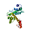
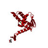
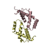
- PDB-3wxa: X-ray crystal structural analysis of the complex between ALG-2 an... -
+ Open data

- Basic information
| Entry | Database: PDB / ID: 3wxa | ||||||
|---|---|---|---|---|---|---|---|
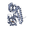
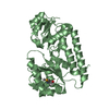
| Title | X-ray crystal structural analysis of the complex between ALG-2 and Sec31A peptide | ||||||
 Components Components |
| ||||||
 Keywords Keywords | APOPTOSIS/TRANSPORT PROTEIN / PENTA-EF-HAND PROTEIN / Endoplasmic reticulum / Membrane / Transport / Apoptosis / Calcium Binding / APOPTOSIS-TRANSPORT PROTEIN complex | ||||||
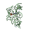
| Function / homology |  Function and homology information Function and homology informationvesicle coat / neural crest formation / positive regulation of ER to Golgi vesicle-mediated transport / vascular endothelial growth factor receptor-2 signaling pathway / COPII-coated vesicle cargo loading / neural crest cell development / COPII vesicle coat assembly / COPII vesicle coat / positive regulation of protein exit from endoplasmic reticulum / XBP1(S) activates chaperone genes ...vesicle coat / neural crest formation / positive regulation of ER to Golgi vesicle-mediated transport / vascular endothelial growth factor receptor-2 signaling pathway / COPII-coated vesicle cargo loading / neural crest cell development / COPII vesicle coat assembly / COPII vesicle coat / positive regulation of protein exit from endoplasmic reticulum / XBP1(S) activates chaperone genes / endoplasmic reticulum organization / COPII-coated ER to Golgi transport vesicle / negative regulation of TOR signaling / COPII-mediated vesicle transport / positive regulation of protein monoubiquitination / negative regulation of vascular endothelial growth factor receptor signaling pathway / Cul3-RING ubiquitin ligase complex / endoplasmic reticulum exit site / endoplasmic reticulum to Golgi vesicle-mediated transport / ubiquitin-like ligase-substrate adaptor activity / protein-membrane adaptor activity / positive regulation of endothelial cell proliferation / MHC class II antigen presentation / positive regulation of endothelial cell migration / negative regulation of phosphatidylinositol 3-kinase/protein kinase B signal transduction / apoptotic signaling pathway / Antigen Presentation: Folding, assembly and peptide loading of class I MHC / ER to Golgi transport vesicle membrane / intracellular protein transport / response to calcium ion / positive regulation of angiogenesis / calcium-dependent protein binding / Signaling by ALK fusions and activated point mutants / cellular response to heat / protein transport / angiogenesis / cytoplasmic vesicle / protein-macromolecule adaptor activity / endosome / protein dimerization activity / positive regulation of apoptotic process / protein heterodimerization activity / calcium ion binding / endoplasmic reticulum membrane / perinuclear region of cytoplasm / structural molecule activity / magnesium ion binding / endoplasmic reticulum / protein homodimerization activity / extracellular exosome / nucleoplasm / identical protein binding / nucleus / cytosol / cytoplasm Similarity search - Function | ||||||
| Biological species |  Homo sapiens (human) Homo sapiens (human) | ||||||
| Method |  X-RAY DIFFRACTION / SYNCHROTRON / MOLECULAR REPLACEMENT / Resolution: 2.36 Å X-RAY DIFFRACTION / SYNCHROTRON / MOLECULAR REPLACEMENT / Resolution: 2.36 Å | ||||||
 Authors Authors | Takahashi, T. / Suzuki, H. / Kawasaki, M. / Shibata, H. / Wakatsuki, S. / Maki, M. | ||||||
 Citation Citation | Journal: Int J Mol Sci / Year: 2015 Title: Structural Analysis of the Complex between Penta-EF-Hand ALG-2 Protein and Sec31A Peptide Reveals a Novel Target Recognition Mechanism of ALG-2 Authors: Takahashi, T. / Kojima, K. / Zhang, W. / Sasaki, K. / Ito, M. / Suzuki, H. / Kawasaki, M. / Wakatsuki, S. / Takahara, T. / Shibata, H. / Maki, M. | ||||||
| History |
|
- Structure visualization
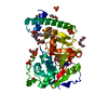
| Structure viewer | Molecule: MolmilJmol/JSmol |
|---|
- Downloads & links
-Download
| PDBx/mmCIF format | 3wxa.cif.gz | 88.3 KB | Display | PDBx/mmCIF format |
|---|---|---|---|---|
| PDB format | pdb3wxa.ent.gz | 66.5 KB | Display | PDB format |
| PDBx/mmJSON format | 3wxa.json.gz | Tree view | PDBx/mmJSON format | |
| Others |  Other downloads Other downloads |
-Validation report
| Arichive directory | https://data.pdbj.org/pub/pdb/validation_reports/wx/3wxaftp://data.pdbj.org/pub/pdb/validation_reports/wx/3wxa | HTTPS FTP |
|---|
-Related structure data
| Related structure data |  2zndS S: Starting model for refinement |
|---|---|
| Similar structure data |
-Links
 PDBj
PDBj


- Assembly
| Deposited unit | 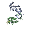
| ||||||||
|---|---|---|---|---|---|---|---|---|---|
| 1 | 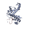
| ||||||||
| 2 | 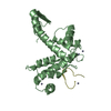
| ||||||||
| Unit cell |
|
-Components
| #1: Protein | Mass: 20158.492 Da / Num. of mol.: 2 / Fragment: UNP residues 20-191 Source method: isolated from a genetically manipulated source Source: (gene. exp.) Homo sapiens (human) / Gene: PDCD6, ALG2 / Plasmid: pET3d / Production host:  #2: Protein/peptide | Mass: 1278.458 Da / Num. of mol.: 2 / Fragment: ALG-2 binding site, UNP residues 837-848 / Source method: obtained synthetically / Details: This sequence occurs naturally in humans. / Source: (synth.) Homo sapiens (human) / References: UniProt: O94979#3: Chemical | ChemComp-ZN /   Mass: 65.409 Da / Num. of mol.: 10 / Source method: obtained synthetically / Formula: Zn Mass: 65.409 Da / Num. of mol.: 10 / Source method: obtained synthetically / Formula: Zn#4: Water | ChemComp-HOH / |  Mass: 18.015 Da / Num. of mol.: 28 / Source method: isolated from a natural source / Formula: H2O Mass: 18.015 Da / Num. of mol.: 28 / Source method: isolated from a natural source / Formula: H2O |
|---|
-Experimental details
-Experiment
| Experiment | Method: X-RAY DIFFRACTION / Number of used crystals: 1 |
|---|
- Sample preparation
| Crystal | Density Matthews: 2.34 Å3/Da / Density % sol: 47.33 % |
|---|---|
| Crystal grow | Temperature: 293 K / Method: vapor diffusion, hanging drop / pH: 6 Details: 0.1M Cacodylate, 20% MPD, 0.05M Zinc Acetate, pH 6.0, VAPOR DIFFUSION, HANGING DROP, temperature 293K |
-Data collection
| Diffraction | Mean temperature: 100 K |
|---|---|
| Diffraction source | Source: SYNCHROTRON / Site: Photon Factory  / Beamline: AR-NE3A / Wavelength: 1.28209 Å / Beamline: AR-NE3A / Wavelength: 1.28209 Å |
| Detector | Type: ADSC QUANTUM 270 / Detector: CCD / Date: Oct 21, 2011 |
| Radiation | Monochromator: Si 111 CHANNEL / Protocol: SINGLE WAVELENGTH / Monochromatic (M) / Laue (L): M / Scattering type: x-ray |
| Radiation wavelength | Wavelength: 1.28209 Å / Relative weight: 1 |
| Reflection | Resolution: 2.36→71 Å / Num. all: 16251 / Num. obs: 16160 / % possible obs: 99.4 % / Redundancy: 10.3 % / Biso Wilson estimate: 36.9 Å2 / Rsym value: 0.08 / Net I/σ(I): 33 |
| Reflection shell | Resolution: 2.36→2.421 Å / Redundancy: 10.3 % / Mean I/σ(I) obs: 33 / Num. unique all: 16251 / Rsym value: 0.08 / % possible all: 99.4 |
- Processing
| Software |
| ||||||||||||||||||||||||||||||||||||||||||
|---|---|---|---|---|---|---|---|---|---|---|---|---|---|---|---|---|---|---|---|---|---|---|---|---|---|---|---|---|---|---|---|---|---|---|---|---|---|---|---|---|---|---|---|
| Refinement | Method to determine structure: MOLECULAR REPLACEMENT Starting model: 2ZND Resolution: 2.36→38.094 Å / σ(F): 1.37 / Phase error: 28.13 / Stereochemistry target values: TWIN_LSQ_F
| ||||||||||||||||||||||||||||||||||||||||||
| Solvent computation | Shrinkage radii: 0.9 Å / VDW probe radii: 1.11 Å / Solvent model: FLAT BULK SOLVENT MODEL | ||||||||||||||||||||||||||||||||||||||||||
| Refinement step | Cycle: LAST / Resolution: 2.36→38.094 Å
| ||||||||||||||||||||||||||||||||||||||||||
| Refine LS restraints |
| ||||||||||||||||||||||||||||||||||||||||||
| LS refinement shell | Refine-ID: X-RAY DIFFRACTION / Total num. of bins used: 6
|
