 Movie
Movie Controller


+ Open data

- Basic information
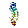
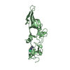
| Entry | Database: PDB / ID: 3wx4 | ||||||
|---|---|---|---|---|---|---|---|
| Title | CRYSTAL STRUCTURE of T4 PHAGE ARN PROTEIN | ||||||
 Components Components | Anti-restriction endonuclease | ||||||
 Keywords Keywords | VIRAL PROTEIN / DNA MIMIC / GENE REGULATION | ||||||
| Function / homology |  Function and homology information Function and homology informationsymbiont-mediated evasion of host restriction-modification system / endonuclease activity / symbiont-mediated suppression of host innate immune response / hydrolase activity Similarity search - Function | ||||||
| Biological species |  Enterobacteria phage T4 (virus) Enterobacteria phage T4 (virus) | ||||||
| Method |  X-RAY DIFFRACTION / SYNCHROTRON / SAD / Resolution: 1.9 Å X-RAY DIFFRACTION / SYNCHROTRON / SAD / Resolution: 1.9 Å | ||||||
 Authors Authors | Ho, C.H. / Wang, H.C. / Ko, T.P. / Wang, A.H.J. | ||||||
 Citation Citation | Journal: J.Biol.Chem. / Year: 2014 Title: The T4 phage DNA mimic protein Arn inhibits the DNA binding activity of the bacterial histone-like protein H-NS Authors: Ho, C.H. / Wang, H.C. / Ko, T.P. / Chang, Y.C. / Wang, A.H.J. | ||||||
| History |
|
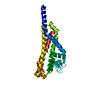
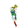
- Structure visualization
| Structure viewer | Molecule: MolmilJmol/JSmol |
|---|
- Downloads & links
-Download
| PDBx/mmCIF format | 3wx4.cif.gz | 35 KB | Display | PDBx/mmCIF format |
|---|---|---|---|---|
| PDB format | pdb3wx4.ent.gz | 24.5 KB | Display | PDB format |
| PDBx/mmJSON format | 3wx4.json.gz | Tree view | PDBx/mmJSON format | |
| Others |  Other downloads Other downloads |
-Validation report
| Arichive directory | https://data.pdbj.org/pub/pdb/validation_reports/wx/3wx4ftp://data.pdbj.org/pub/pdb/validation_reports/wx/3wx4 | HTTPS FTP |
|---|
-Related structure data
| Similar structure data |
|---|


-Links
 PDBj
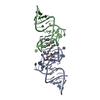
PDBj- Assembly
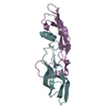
| Deposited unit | 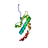
| ||||||||||||
|---|---|---|---|---|---|---|---|---|---|---|---|---|---|
| 1 | 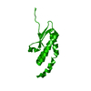
| ||||||||||||
| Unit cell |
| ||||||||||||
| Components on special symmetry positions |
|
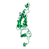
-Components
| #1: Protein | Mass: 11981.584 Da / Num. of mol.: 1 Source method: isolated from a genetically manipulated source Source: (gene. exp.) Enterobacteria phage T4 (virus) / Gene: arn, asiA.1, motA.-6 / Plasmid: PET21B / Production host:  |
|---|---|
| #2: Water | ChemComp-HOH /  Mass: 18.015 Da / Num. of mol.: 155 / Source method: isolated from a natural source / Formula: H2O Mass: 18.015 Da / Num. of mol.: 155 / Source method: isolated from a natural source / Formula: H2O |
-Experimental details
-Experiment
| Experiment | Method: X-RAY DIFFRACTION / Number of used crystals: 1 |
|---|
- Sample preparation
| Crystal | Density Matthews: 4.03 Å3/Da / Density % sol: 69.48 % |
|---|---|
| Crystal grow | pH: 8 Details: 50MM TRIS, 100MM NACL, 3.6M NA- FORMATE(RESERVOIR), 0.1M TRIS(RESERVOIR), PH 8.0, VAPOR DIFFUSION, SITTING DROP, TEMPERATURE 298K |
-Data collection
| Diffraction | Mean temperature: 100 K |
|---|---|
| Diffraction source | Source: SYNCHROTRON / Site: NSRRC  / Beamline: BL13B1 / Wavelength: 0.9762 / Beamline: BL13B1 / Wavelength: 0.9762 |
| Detector | Type: ADSC QUANTUM 315r / Detector: CCD / Date: Nov 4, 2010 / Details: RH COATED MIRRORS |
| Radiation | Monochromator: SI(111) / Protocol: SINGLE WAVELENGTH / Monochromatic (M) / Laue (L): M / Scattering type: x-ray |
| Radiation wavelength | Wavelength: 0.9762 Å / Relative weight: 1 |
| Reflection | Resolution: 1.9→25 Å / Num. obs: 15419 / % possible obs: 99.4 % / Observed criterion σ(I): 5 |
| Reflection shell | Resolution: 1.9→1.97 Å / Rmerge(I) obs: 0.478 / Mean I/σ(I) obs: 6.15 / % possible all: 100 |
- Processing
| Software |
| ||||||||||||||||||||||||||||||||||||||||||||||||||||||||||||
|---|---|---|---|---|---|---|---|---|---|---|---|---|---|---|---|---|---|---|---|---|---|---|---|---|---|---|---|---|---|---|---|---|---|---|---|---|---|---|---|---|---|---|---|---|---|---|---|---|---|---|---|---|---|---|---|---|---|---|---|---|---|
| Refinement | Method to determine structure: SAD / Resolution: 1.9→24 Å / σ(F): 0 / Stereochemistry target values: ENGH & HUBER
| ||||||||||||||||||||||||||||||||||||||||||||||||||||||||||||
| Refine analyze |
| ||||||||||||||||||||||||||||||||||||||||||||||||||||||||||||
| Refinement step | Cycle: LAST / Resolution: 1.9→24 Å
| ||||||||||||||||||||||||||||||||||||||||||||||||||||||||||||
| Refine LS restraints |
|
