 Movie
Movie Controller


+ Open data

- Basic information
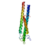
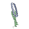
| Entry | Database: PDB / ID: 3wut | ||||||
|---|---|---|---|---|---|---|---|
| Title | Structure basis of inactivating cell abscission | ||||||
 Components Components |
| ||||||
 Keywords Keywords | CELL CYCLE / Coiled-coil | ||||||
| Function / homology |  Function and homology information Function and homology informationintercellular bridge organization / negative regulation of cytokinesis / mitotic sister chromatid separation / attachment of spindle microtubules to kinetochore / regulation of phosphatidylinositol 3-kinase/protein kinase B signal transduction / male meiotic nuclear division / midbody abscission / cranial skeletal system development / Flemming body / mitotic spindle assembly checkpoint signaling ...intercellular bridge organization / negative regulation of cytokinesis / mitotic sister chromatid separation / attachment of spindle microtubules to kinetochore / regulation of phosphatidylinositol 3-kinase/protein kinase B signal transduction / male meiotic nuclear division / midbody abscission / cranial skeletal system development / Flemming body / mitotic spindle assembly checkpoint signaling / cleavage furrow / mitotic cytokinesis / intercellular bridge / cellular response to leukemia inhibitory factor / establishment of protein localization / centriole / kinetochore / midbody / protein kinase activity / cell division / centrosome / protein kinase binding / extracellular exosome / ATP binding / membrane / identical protein binding / cytoplasm Similarity search - Function | ||||||
| Biological species |  Homo sapiens (human) Homo sapiens (human) | ||||||
| Method |  X-RAY DIFFRACTION / SYNCHROTRON / MOLECULAR REPLACEMENT / Resolution: 2.301 Å X-RAY DIFFRACTION / SYNCHROTRON / MOLECULAR REPLACEMENT / Resolution: 2.301 Å | ||||||
 Authors Authors | Kim, H.J. / Matsuura, A. / Lee, H.H. | ||||||
 Citation Citation | Journal: Proc.Natl.Acad.Sci.USA / Year: 2015 Title: Structural and biochemical insights into the role of testis-expressed gene 14 (TEX14) in forming the stable intercellular bridges of germ cells. Authors: Kim, H.J. / Yoon, J. / Matsuura, A. / Na, J.H. / Lee, W.K. / Kim, H. / Choi, J.W. / Park, J.E. / Park, S.J. / Kim, K.T. / Chang, R. / Lee, B.I. / Yu, Y.G. / Shin, Y.K. / Jeong, C. / Rhee, K. / Lee, H.H. | ||||||
| History |
|
- Structure visualization
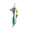
| Structure viewer | Molecule: MolmilJmol/JSmol |
|---|
- Downloads & links
-Download
| PDBx/mmCIF format | 3wut.cif.gz | 106.3 KB | Display | PDBx/mmCIF format |
|---|---|---|---|---|
| PDB format | pdb3wut.ent.gz | 83.9 KB | Display | PDB format |
| PDBx/mmJSON format | 3wut.json.gz | Tree view | PDBx/mmJSON format | |
| Others |  Other downloads Other downloads |
-Validation report
| Arichive directory | https://data.pdbj.org/pub/pdb/validation_reports/wu/3wutftp://data.pdbj.org/pub/pdb/validation_reports/wu/3wut | HTTPS FTP |
|---|
-Related structure data


-Links
 PDBj
PDBj- Assembly
| Deposited unit | 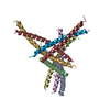
| ||||||||
|---|---|---|---|---|---|---|---|---|---|
| 1 | 
| ||||||||
| 2 | 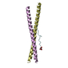
| ||||||||
| 3 | 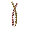
| ||||||||
| 4 | 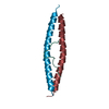
| ||||||||
| Unit cell |
|
-Components
| #1: Protein | Mass: 7243.210 Da / Num. of mol.: 8 / Fragment: UNP residues 160-217 Source method: isolated from a genetically manipulated source Source: (gene. exp.) Homo sapiens (human) / Gene: CEP55 / Plasmid: pGST2 / Production host:  #2: Protein/peptide | Mass: 1452.650 Da / Num. of mol.: 4 / Fragment: UNP residues 792-804 / Source method: obtained synthetically / Details: This sequence occurs naturally in humans. / Source: (synth.) Homo sapiens (human) / References: UniProt: Q8IWB6#3: Chemical |   Mass: 92.094 Da / Num. of mol.: 2 / Source method: obtained synthetically / Formula: C3H8O3 Mass: 92.094 Da / Num. of mol.: 2 / Source method: obtained synthetically / Formula: C3H8O3#4: Water | ChemComp-HOH / |  Mass: 18.015 Da / Num. of mol.: 188 / Source method: isolated from a natural source / Formula: H2O Mass: 18.015 Da / Num. of mol.: 188 / Source method: isolated from a natural source / Formula: H2O |
|---|
-Experimental details
-Experiment
| Experiment | Method: X-RAY DIFFRACTION / Number of used crystals: 1 |
|---|
- Sample preparation
| Crystal | Density Matthews: 2.88 Å3/Da / Density % sol: 57.22 % |
|---|---|
| Crystal grow | Temperature: 295 K / Method: vapor diffusion, sitting drop / pH: 8 Details: 1M ammonium phosphate dibasic, pH 8.0, VAPOR DIFFUSION, SITTING DROP, temperature 295K |
-Data collection
| Diffraction | Mean temperature: 100 K |
|---|---|
| Diffraction source | Source: SYNCHROTRON / Site: PAL/PLS  / Beamline: 7A (6B, 6C1) / Wavelength: 0.97928 Å / Beamline: 7A (6B, 6C1) / Wavelength: 0.97928 Å |
| Detector | Type: ADSC QUANTUM 270 / Detector: CCD / Date: Nov 3, 2012 |
| Radiation | Monochromator: Si 111 DCM / Protocol: SINGLE WAVELENGTH / Monochromatic (M) / Laue (L): M / Scattering type: x-ray |
| Radiation wavelength | Wavelength: 0.97928 Å / Relative weight: 1 |
| Reflection | Resolution: 2.3→30 Å / Num. all: 34110 / Num. obs: 31519 / % possible obs: 99.6 % / Observed criterion σ(F): 0 / Observed criterion σ(I): -3 / Biso Wilson estimate: 43.14 Å2 |
| Reflection shell | Highest resolution: 2.3 Å / % possible all: 99.6 |
- Processing
| Software |
| ||||||||||||||||||||||||||||||||||||||||||||||||||||||||||||||||||||||||||||||||||||
|---|---|---|---|---|---|---|---|---|---|---|---|---|---|---|---|---|---|---|---|---|---|---|---|---|---|---|---|---|---|---|---|---|---|---|---|---|---|---|---|---|---|---|---|---|---|---|---|---|---|---|---|---|---|---|---|---|---|---|---|---|---|---|---|---|---|---|---|---|---|---|---|---|---|---|---|---|---|---|---|---|---|---|---|---|---|
| Refinement | Method to determine structure: MOLECULAR REPLACEMENT / Resolution: 2.301→28.884 Å / FOM work R set: 0.801 / SU ML: 0.25 / σ(F): 0.16 / Phase error: 26.49 / Stereochemistry target values: ML
| ||||||||||||||||||||||||||||||||||||||||||||||||||||||||||||||||||||||||||||||||||||
| Solvent computation | Shrinkage radii: 0.9 Å / VDW probe radii: 1.11 Å / Solvent model: FLAT BULK SOLVENT MODEL | ||||||||||||||||||||||||||||||||||||||||||||||||||||||||||||||||||||||||||||||||||||
| Displacement parameters | Biso max: 134.37 Å2 / Biso mean: 61.47 Å2 / Biso min: 31.28 Å2 | ||||||||||||||||||||||||||||||||||||||||||||||||||||||||||||||||||||||||||||||||||||
| Refinement step | Cycle: LAST / Resolution: 2.301→28.884 Å
| ||||||||||||||||||||||||||||||||||||||||||||||||||||||||||||||||||||||||||||||||||||
| Refine LS restraints |
| ||||||||||||||||||||||||||||||||||||||||||||||||||||||||||||||||||||||||||||||||||||
| LS refinement shell | Refine-ID: X-RAY DIFFRACTION / Total num. of bins used: 11
|
