 Movie
Movie Controller

[English] 日本語
 Yorodumi
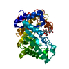
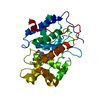
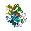
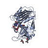
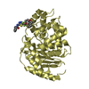
- PDB-3wug: The mutant crystal structure of b-1,4-Xylanase (XynAS9_V43P/G44E)... -
+ Open data

- Basic information
| Entry | Database: PDB / ID: 3wug | |||||||||
|---|---|---|---|---|---|---|---|---|---|---|
| Title | The mutant crystal structure of b-1,4-Xylanase (XynAS9_V43P/G44E) with xylobiose from Streptomyces sp. 9 | |||||||||
 Components Components | Endo-1,4-beta-xylanase A | |||||||||
 Keywords Keywords | HYDROLASE / beta-1 / 4-xylanase / thermozyme / protein engineering / protein rigidity | |||||||||
| Function / homology |  Function and homology information Function and homology informationendo-1,4-beta-xylanase / endo-1,4-beta-xylanase activity / xylan catabolic process / polysaccharide binding Similarity search - Function | |||||||||
| Biological species |  Streptomyces sp. (bacteria) Streptomyces sp. (bacteria) | |||||||||
| Method |  X-RAY DIFFRACTION / SYNCHROTRON / MOLECULAR REPLACEMENT / Resolution: 1.88 Å X-RAY DIFFRACTION / SYNCHROTRON / MOLECULAR REPLACEMENT / Resolution: 1.88 Å | |||||||||
 Authors Authors | Chen, C.C. / Han, X. / Lv, P. / Ko, T.P. / Peng, W. / Huang, C.H. / Zheng, Y. / Gao, J. / Yang, Y.Y. / Guo, R.T. | |||||||||
 Citation Citation | Journal: J.Biotechnol. / Year: 2014 Title: Structural perspectives of an engineered beta-1,4-xylanase with enhanced thermostability. Authors: Chen, C.C. / Luo, H. / Han, X. / Lv, P. / Ko, T.P. / Peng, W. / Huang, C.H. / Wang, K. / Gao, J. / Zheng, Y. / Yang, Y. / Zhang, J. / Yao, B. / Guo, R.T. | |||||||||
| History |
|
- Structure visualization
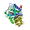
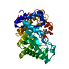
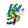
| Structure viewer | Molecule: MolmilJmol/JSmol |
|---|
- Downloads & links
-Download
| PDBx/mmCIF format | 3wug.cif.gz | 84 KB | Display | PDBx/mmCIF format |
|---|---|---|---|---|
| PDB format | pdb3wug.ent.gz | 61.2 KB | Display | PDB format |
| PDBx/mmJSON format | 3wug.json.gz | Tree view | PDBx/mmJSON format | |
| Others |  Other downloads Other downloads |
-Validation report
| Arichive directory | https://data.pdbj.org/pub/pdb/validation_reports/wu/3wugftp://data.pdbj.org/pub/pdb/validation_reports/wu/3wug | HTTPS FTP |
|---|
-Related structure data
| Related structure data |  3wubC 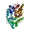 3wueC  3wufC  3cuiS C: citing same article ( S: Starting model for refinement |
|---|---|
| Similar structure data |

-Links
 PDBj
PDBj

- Assembly
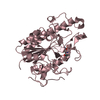
| Deposited unit | 
| ||||||||
|---|---|---|---|---|---|---|---|---|---|
| 1 |
| ||||||||
| Unit cell |
|
-Components
| #1: Protein | Mass: 34681.492 Da / Num. of mol.: 1 / Fragment: UNP RESIDUES 39-351 / Mutation: V81P, G82E Source method: isolated from a genetically manipulated source Source: (gene. exp.) Streptomyces sp. (bacteria) / Gene: xynAS9 / Plasmid: pPIC9 / Production host:  Komagataella pastoris (fungus) / Strain (production host): GS115 / References: UniProt: B4XVN1, endo-1,4-beta-xylanase Komagataella pastoris (fungus) / Strain (production host): GS115 / References: UniProt: B4XVN1, endo-1,4-beta-xylanase | ||||||
|---|---|---|---|---|---|---|---|
| #2: Polysaccharide | 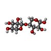  Source method: isolated from a genetically manipulated source Details: oligosaccharide / References: 4beta-beta-xylobiose #3: Chemical | ChemComp-ZN /   Mass: 65.409 Da / Num. of mol.: 7 / Source method: obtained synthetically / Formula: Zn Mass: 65.409 Da / Num. of mol.: 7 / Source method: obtained synthetically / Formula: Zn#4: Water | ChemComp-HOH / |  Mass: 18.015 Da / Num. of mol.: 316 / Source method: isolated from a natural source / Formula: H2O Mass: 18.015 Da / Num. of mol.: 316 / Source method: isolated from a natural source / Formula: H2OHas protein modification | Y | |
-Experimental details
-Experiment
| Experiment | Method: X-RAY DIFFRACTION / Number of used crystals: 1 |
|---|
- Sample preparation
| Crystal | Density Matthews: 3.92 Å3/Da / Density % sol: 68.63 % |
|---|---|
| Crystal grow | Temperature: 298 K / Method: vapor diffusion, sitting drop / pH: 6.5 Details: 0.5M zinc acetate dehydrate, 0.1M sodium cacodylate trihydrate pH 6.5, 8% (w/v) , VAPOR DIFFUSION, SITTING DROP, temperature 298K |
-Data collection
| Diffraction | Mean temperature: 100 K |
|---|---|
| Diffraction source | Source: SYNCHROTRON / Site: NSRRC  / Beamline: BL13C1 / Wavelength: 1 Å / Beamline: BL13C1 / Wavelength: 1 Å |
| Detector | Type: ADSC QUANTUM 315 / Detector: CCD / Date: Jul 20, 2013 |
| Radiation | Protocol: SINGLE WAVELENGTH / Monochromatic (M) / Laue (L): M / Scattering type: x-ray |
| Radiation wavelength | Wavelength: 1 Å / Relative weight: 1 |
| Reflection | Resolution: 1.88→25 Å / Num. obs: 46576 / % possible obs: 99.9 % / Redundancy: 7.7 % / Rmerge(I) obs: 0.077 / Net I/σ(I): 32 |
| Reflection shell | Resolution: 1.88→1.95 Å / Redundancy: 7.8 % / Rmerge(I) obs: 0.477 / Mean I/σ(I) obs: 4.5 / % possible all: 100 |
- Processing
| Software |
| ||||||||||||||||||||
|---|---|---|---|---|---|---|---|---|---|---|---|---|---|---|---|---|---|---|---|---|---|
| Refinement | Method to determine structure: MOLECULAR REPLACEMENT Starting model: 3CUI Resolution: 1.88→25 Å / Stereochemistry target values: Engh & Huber
| ||||||||||||||||||||
| Refinement step | Cycle: LAST / Resolution: 1.88→25 Å
| ||||||||||||||||||||
| LS refinement shell | Resolution: 1.88→1.95 Å /
|
