 Movie
Movie Controller

[English] 日本語
 Yorodumi
- PDB-3wpx: Structure of PomBc4, a periplasmic fragment of PomB from Vibrio a... -
+ Open data

- Basic information
| Entry | Database: PDB / ID: 3wpx | ||||||
|---|---|---|---|---|---|---|---|
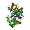
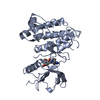
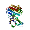
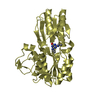
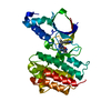
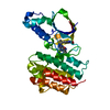
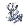
| Title | Structure of PomBc4, a periplasmic fragment of PomB from Vibrio alginolyticus | ||||||
 Components Components | PomB | ||||||
 Keywords Keywords | MEMBRANE PROTEIN / ompA-like / stator / peptidoglycan | ||||||
| Function / homology |  Function and homology information Function and homology information | ||||||
| Biological species |  Vibrio alginolyticus (bacteria) Vibrio alginolyticus (bacteria) | ||||||
| Method |  X-RAY DIFFRACTION / SYNCHROTRON / MOLECULAR REPLACEMENT / Resolution: 2.3 Å X-RAY DIFFRACTION / SYNCHROTRON / MOLECULAR REPLACEMENT / Resolution: 2.3 Å | ||||||
 Authors Authors | Takao, M. / Sakuma, M. / Zhu, S. / Homma, M. / Kojima, S. / Imada, K. | ||||||
 Citation Citation | Journal: Proc. Natl. Acad. Sci. U.S.A. / Year: 2014 Title: Conformational change in the periplasmic region of the flagellar stator coupled with the assembly around the rotor Authors: Zhu, S. / Takao, M. / Li, N. / Sakuma, M. / Nishino, Y. / Homma, M. / Kojima, S. / Imada, K. | ||||||
| History |
|
- Structure visualization
| Structure viewer | Molecule: MolmilJmol/JSmol |
|---|
- Downloads & links
-Download
| PDBx/mmCIF format | 3wpx.cif.gz | 74.2 KB | Display | PDBx/mmCIF format |
|---|---|---|---|---|
| PDB format | pdb3wpx.ent.gz | 54.8 KB | Display | PDB format |
| PDBx/mmJSON format | 3wpx.json.gz | Tree view | PDBx/mmJSON format | |
| Others |  Other downloads Other downloads |
-Validation report
| Arichive directory | https://data.pdbj.org/pub/pdb/validation_reports/wp/3wpxftp://data.pdbj.org/pub/pdb/validation_reports/wp/3wpx | HTTPS FTP |
|---|
-Related structure data
| Related structure data |  3wpwSC S: Starting model for refinement C: citing same article ( |
|---|---|
| Similar structure data |


-Links
 PDBj
PDBj- Assembly
| Deposited unit | 
| ||||||||
|---|---|---|---|---|---|---|---|---|---|
| 1 |
| ||||||||
| Unit cell |
|
-Components
| #1: Protein | Mass: 22846.504 Da / Num. of mol.: 2 / Fragment: UNP residues 121-315 Source method: isolated from a genetically manipulated source Source: (gene. exp.) Vibrio alginolyticus (bacteria) / Gene: pomB / Plasmid: pET19b / Production host: #2: Water | ChemComp-HOH / |  Mass: 18.015 Da / Num. of mol.: 57 / Source method: isolated from a natural source / Formula: H2O Mass: 18.015 Da / Num. of mol.: 57 / Source method: isolated from a natural source / Formula: H2O |
|---|
-Experimental details
-Experiment
| Experiment | Method: X-RAY DIFFRACTION / Number of used crystals: 1 |
|---|
- Sample preparation
| Crystal | Density Matthews: 1.56 Å3/Da / Density % sol: 21.25 % |
|---|---|
| Crystal grow | Temperature: 293 K / Method: vapor diffusion, sitting drop / pH: 6.5 Details: 20%(w/v) PEGMME2000, 0.1M imidazole, 4%(v/v) MPD, pH 6.5, VAPOR DIFFUSION, SITTING DROP, temperature 293K |
-Data collection
| Diffraction | Mean temperature: 100 K |
|---|---|
| Diffraction source | Source: SYNCHROTRON / Site: SPring-8  / Beamline: BL32XU / Wavelength: 0.9791 Å / Beamline: BL32XU / Wavelength: 0.9791 Å |
| Detector | Type: RAYONIX MX225HE / Detector: CCD / Date: Feb 16, 2011 |
| Radiation | Monochromator: Double-crystal monochromator / Protocol: SINGLE WAVELENGTH / Monochromatic (M) / Laue (L): M / Scattering type: x-ray |
| Radiation wavelength | Wavelength: 0.9791 Å / Relative weight: 1 |
| Reflection | Resolution: 2.3→38.6 Å / Num. all: 12786 / Num. obs: 12786 / % possible obs: 97.2 % / Observed criterion σ(F): 0 / Observed criterion σ(I): 0 / Redundancy: 4.6 % / Biso Wilson estimate: 36.6 Å2 / Rmerge(I) obs: 0.099 / Net I/σ(I): 9.7 |
| Reflection shell | Resolution: 2.3→2.42 Å / Redundancy: 4.6 % / Rmerge(I) obs: 0.391 / Mean I/σ(I) obs: 3.7 / Num. unique all: 1876 / % possible all: 98.5 |
- Processing
| Software |
| ||||||||||||||||||||||||||||||||||||||||||||||||||||||||||||
|---|---|---|---|---|---|---|---|---|---|---|---|---|---|---|---|---|---|---|---|---|---|---|---|---|---|---|---|---|---|---|---|---|---|---|---|---|---|---|---|---|---|---|---|---|---|---|---|---|---|---|---|---|---|---|---|---|---|---|---|---|---|
| Refinement | Method to determine structure: MOLECULAR REPLACEMENT Starting model: 3WPW Resolution: 2.3→32.96 Å / SU ML: 0.63 / σ(F): 1.34 / Phase error: 23.38 / Stereochemistry target values: ML
| ||||||||||||||||||||||||||||||||||||||||||||||||||||||||||||
| Solvent computation | Shrinkage radii: 0.83 Å / VDW probe radii: 1.1 Å / Solvent model: FLAT BULK SOLVENT MODEL / Bsol: 44.65 Å2 / ksol: 0.351 e/Å3 | ||||||||||||||||||||||||||||||||||||||||||||||||||||||||||||
| Displacement parameters |
| ||||||||||||||||||||||||||||||||||||||||||||||||||||||||||||
| Refinement step | Cycle: LAST / Resolution: 2.3→32.96 Å
| ||||||||||||||||||||||||||||||||||||||||||||||||||||||||||||
| Refine LS restraints |
| ||||||||||||||||||||||||||||||||||||||||||||||||||||||||||||
| LS refinement shell | Refine-ID: X-RAY DIFFRACTION / Total num. of bins used: 9
|
