 Movie
Movie Controller


+ Open data

- Basic information
| Entry | Database: PDB / ID: 3woq | ||||||
|---|---|---|---|---|---|---|---|
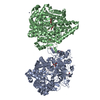
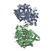
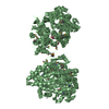
| Title | Crystal structure of the DAP BII hexapeptide complex III | ||||||
 Components Components |
| ||||||
 Keywords Keywords | Hydrolase/Hormone / Chymotrypsin fold / S46 peptidase / Hydrolase-Hormone complex | ||||||
| Function / homology |  Function and homology information Function and homology informationHydrolases; Acting on peptide bonds (peptidases); Dipeptidyl-peptidases and tripeptidyl-peptidases / serine-type aminopeptidase activity / dipeptidyl-peptidase activity / : / protein homodimerization activity / identical protein binding Similarity search - Function | ||||||
| Biological species |  Pseudoxanthomonas mexicana (bacteria) Pseudoxanthomonas mexicana (bacteria) Homo sapiens (human) Homo sapiens (human) | ||||||
| Method |  X-RAY DIFFRACTION / SYNCHROTRON / MOLECULAR REPLACEMENT / Resolution: 1.82 Å X-RAY DIFFRACTION / SYNCHROTRON / MOLECULAR REPLACEMENT / Resolution: 1.82 Å | ||||||
 Authors Authors | Sakamoto, Y. / Suzuki, Y. / Iizuka, I. / Tateoka, C. / Roppongi, S. / Fujimoto, M. / Nonaka, T. / Ogasawara, W. / Tanaka, N. | ||||||
 Citation Citation | Journal: SCI REP / Year: 2014 Title: S46 peptidases are the first exopeptidases to be members of clan PA Authors: Sakamoto, Y. / Suzuki, Y. / Iizuka, I. / Tateoka, C. / Roppongi, S. / Fujimoto, M. / Inaka, K. / Tanaka, H. / Masaki, M. / Ohta, K. / Okada, H. / Nonaka, T. / Morikawa, Y. / Nakamura, K.T. / ...Authors: Sakamoto, Y. / Suzuki, Y. / Iizuka, I. / Tateoka, C. / Roppongi, S. / Fujimoto, M. / Inaka, K. / Tanaka, H. / Masaki, M. / Ohta, K. / Okada, H. / Nonaka, T. / Morikawa, Y. / Nakamura, K.T. / Ogasawara, W. / Tanaka, N. | ||||||
| History |
|
- Structure visualization
| Structure viewer | Molecule: MolmilJmol/JSmol |
|---|
- Downloads & links
-Download
| PDBx/mmCIF format | 3woq.cif.gz | 296.7 KB | Display | PDBx/mmCIF format |
|---|---|---|---|---|
| PDB format | pdb3woq.ent.gz | 236.4 KB | Display | PDB format |
| PDBx/mmJSON format | 3woq.json.gz | Tree view | PDBx/mmJSON format | |
| Others |  Other downloads Other downloads |
-Validation report
| Arichive directory | https://data.pdbj.org/pub/pdb/validation_reports/wo/3woqftp://data.pdbj.org/pub/pdb/validation_reports/wo/3woq | HTTPS FTP |
|---|
-Related structure data
| Related structure data |  3woiSC  3wojC  3wokC  3wolC  3womC  3wonC  3wooC  3wopC  3worC S: Starting model for refinement C: citing same article ( |
|---|---|
| Similar structure data |


-Links
 PDBj
PDBj- Assembly
| Deposited unit | 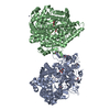
| ||||||||
|---|---|---|---|---|---|---|---|---|---|
| 1 |
| ||||||||
| Unit cell |
|
-Components
| #1: Protein | Mass: 76261.203 Da / Num. of mol.: 2 / Mutation: H86A, D224A, S657A Source method: isolated from a genetically manipulated source Source: (gene. exp.) Pseudoxanthomonas mexicana (bacteria) / Strain: WO24 / Production host: References: UniProt: V5YM14, Hydrolases; Acting on peptide bonds (peptidases); Dipeptidyl-peptidases and tripeptidyl-peptidases #2: Protein/peptide | Mass: 775.913 Da / Num. of mol.: 2 / Source method: obtained synthetically / Details: synthetic peptide / Source: (synth.) Homo sapiens (human)#3: Chemical | ChemComp-GOL /   Mass: 92.094 Da / Num. of mol.: 9 / Source method: obtained synthetically / Formula: C3H8O3 Mass: 92.094 Da / Num. of mol.: 9 / Source method: obtained synthetically / Formula: C3H8O3#4: Chemical | ChemComp-ZN /   Mass: 65.409 Da / Num. of mol.: 4 / Source method: obtained synthetically / Formula: Zn Mass: 65.409 Da / Num. of mol.: 4 / Source method: obtained synthetically / Formula: Zn#5: Water | ChemComp-HOH / |  Mass: 18.015 Da / Num. of mol.: 775 / Source method: isolated from a natural source / Formula: H2O Mass: 18.015 Da / Num. of mol.: 775 / Source method: isolated from a natural source / Formula: H2OHas protein modification | Y | |
|---|
-Experimental details
-Experiment
| Experiment | Method: X-RAY DIFFRACTION / Number of used crystals: 1 |
|---|
- Sample preparation
| Crystal | Density Matthews: 2.55 Å3/Da / Density % sol: 51.76 % |
|---|---|
| Crystal grow | Temperature: 293 K / Method: vapor diffusion, hanging drop / pH: 9.5 Details: 18% PEG 8000, 20% Glycerol, 2mM ZnCl2, 80mM CHES, 2mM Angiotensin IV, pH 9.5, VAPOR DIFFUSION, HANGING DROP, temperature 293K |
-Data collection
| Diffraction | Mean temperature: 95 K |
|---|---|
| Diffraction source | Source: SYNCHROTRON / Site: Photon Factory  / Beamline: BL-17A / Wavelength: 0.98 Å / Beamline: BL-17A / Wavelength: 0.98 Å |
| Detector | Type: ADSC QUANTUM 270 / Detector: CCD / Date: Jan 27, 2013 |
| Radiation | Protocol: SINGLE WAVELENGTH / Monochromatic (M) / Laue (L): M / Scattering type: x-ray |
| Radiation wavelength | Wavelength: 0.98 Å / Relative weight: 1 |
| Reflection | Resolution: 1.82→40 Å / Num. obs: 138595 / % possible obs: 99.4 % |
- Processing
| Software |
| ||||||||||||||||||||||||||||||||||||||||||||||||||||||||||||||||||||||||||||||||||||||||||||||||||||||||||||||||||||||||||||||||||||||||||||||||||||||||||||||||||||||||||||||||||||||
|---|---|---|---|---|---|---|---|---|---|---|---|---|---|---|---|---|---|---|---|---|---|---|---|---|---|---|---|---|---|---|---|---|---|---|---|---|---|---|---|---|---|---|---|---|---|---|---|---|---|---|---|---|---|---|---|---|---|---|---|---|---|---|---|---|---|---|---|---|---|---|---|---|---|---|---|---|---|---|---|---|---|---|---|---|---|---|---|---|---|---|---|---|---|---|---|---|---|---|---|---|---|---|---|---|---|---|---|---|---|---|---|---|---|---|---|---|---|---|---|---|---|---|---|---|---|---|---|---|---|---|---|---|---|---|---|---|---|---|---|---|---|---|---|---|---|---|---|---|---|---|---|---|---|---|---|---|---|---|---|---|---|---|---|---|---|---|---|---|---|---|---|---|---|---|---|---|---|---|---|---|---|---|---|
| Refinement | Method to determine structure: MOLECULAR REPLACEMENT Starting model: 3WOI Resolution: 1.82→39.92 Å / Cor.coef. Fo:Fc: 0.957 / Cor.coef. Fo:Fc free: 0.938 / SU B: 2.772 / SU ML: 0.085 / Cross valid method: THROUGHOUT / ESU R: 0.122 / ESU R Free: 0.122 / Stereochemistry target values: MAXIMUM LIKELIHOOD / Details: HYDROGENS HAVE BEEN ADDED IN THE RIDING POSITIONS
| ||||||||||||||||||||||||||||||||||||||||||||||||||||||||||||||||||||||||||||||||||||||||||||||||||||||||||||||||||||||||||||||||||||||||||||||||||||||||||||||||||||||||||||||||||||||
| Solvent computation | Ion probe radii: 0.8 Å / Shrinkage radii: 0.8 Å / VDW probe radii: 1.2 Å / Solvent model: MASK | ||||||||||||||||||||||||||||||||||||||||||||||||||||||||||||||||||||||||||||||||||||||||||||||||||||||||||||||||||||||||||||||||||||||||||||||||||||||||||||||||||||||||||||||||||||||
| Displacement parameters | Biso mean: 26.943 Å2
| ||||||||||||||||||||||||||||||||||||||||||||||||||||||||||||||||||||||||||||||||||||||||||||||||||||||||||||||||||||||||||||||||||||||||||||||||||||||||||||||||||||||||||||||||||||||
| Refinement step | Cycle: LAST / Resolution: 1.82→39.92 Å
| ||||||||||||||||||||||||||||||||||||||||||||||||||||||||||||||||||||||||||||||||||||||||||||||||||||||||||||||||||||||||||||||||||||||||||||||||||||||||||||||||||||||||||||||||||||||
| Refine LS restraints |
|