 Movie
Movie Controller


+ Open data

- Basic information
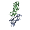
| Entry | Database: PDB / ID: 3wmj | ||||||
|---|---|---|---|---|---|---|---|
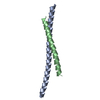
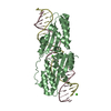
| Title | Crystal structure of EIAV vaccine gp45 | ||||||
 Components Components | (EIAV vaccine gp45) x 2 | ||||||
 Keywords Keywords | VIRAL PROTEIN / alpha-helix / virus fusion / envelope | ||||||
| Function / homology |  Function and homology information Function and homology informationviral envelope / symbiont entry into host cell / virion attachment to host cell / host cell plasma membrane / virion membrane / structural molecule activity Similarity search - Function | ||||||
| Biological species |  | ||||||
| Method |  X-RAY DIFFRACTION / SYNCHROTRON / MOLECULAR REPLACEMENT / Resolution: 1.998 Å X-RAY DIFFRACTION / SYNCHROTRON / MOLECULAR REPLACEMENT / Resolution: 1.998 Å | ||||||
 Authors Authors | Liu, X. / Du, J. / Qiao, W. | ||||||
 Citation Citation | Journal: To be Published Title: A mutation associated with EIAV vaccine strain within heptad repeat of EIAV gp45 provides insight into vaccine development for HIV Authors: Liu, X. / Du, J. / Qiao, W. | ||||||
| History |
|
- Structure visualization
| Structure viewer | Molecule: MolmilJmol/JSmol |
|---|
- Downloads & links
-Download
| PDBx/mmCIF format | 3wmj.cif.gz | 31.5 KB | Display | PDBx/mmCIF format |
|---|---|---|---|---|
| PDB format | pdb3wmj.ent.gz | 21.1 KB | Display | PDB format |
| PDBx/mmJSON format | 3wmj.json.gz | Tree view | PDBx/mmJSON format | |
| Others |  Other downloads Other downloads |
-Validation report
| Arichive directory | https://data.pdbj.org/pub/pdb/validation_reports/wm/3wmjftp://data.pdbj.org/pub/pdb/validation_reports/wm/3wmj | HTTPS FTP |
|---|
-Related structure data


-Links
 PDBj
PDBj


- Assembly
| Deposited unit | 
| ||||||||||||||||||||||||
|---|---|---|---|---|---|---|---|---|---|---|---|---|---|---|---|---|---|---|---|---|---|---|---|---|---|
| 1 | 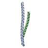
| ||||||||||||||||||||||||
| Unit cell |
| ||||||||||||||||||||||||
| Components on special symmetry positions |
|
-Components
| #1: Protein | Mass: 6580.127 Da / Num. of mol.: 1 Source method: isolated from a genetically manipulated source Source: (gene. exp.)  |
|---|---|
| #2: Protein/peptide | Mass: 4450.874 Da / Num. of mol.: 1 Source method: isolated from a genetically manipulated source Source: (gene. exp.) |
| #3: Water | ChemComp-HOH /  Mass: 18.015 Da / Num. of mol.: 64 / Source method: isolated from a natural source / Formula: H2O Mass: 18.015 Da / Num. of mol.: 64 / Source method: isolated from a natural source / Formula: H2O |
| Sequence details | THE SEQUENCE OF THIS PROTEIN WAS NOT AVAILABLE AT THE UNIPROT KNOWLEDGEBASE DATABASE (UNIPROTKB) AT ...THE SEQUENCE OF THIS PROTEIN WAS NOT AVAILABLE AT THE UNIPROT KNOWLEDGEB |
-Experimental details
-Experiment
| Experiment | Method: X-RAY DIFFRACTION / Number of used crystals: 1 |
|---|
- Sample preparation
| Crystal | Density Matthews: 2.93 Å3/Da / Density % sol: 58.03 % |
|---|---|
| Crystal grow | Temperature: 293 K / Method: evaporation / pH: 4.6 Details: 0.2M NaCl, 0.1M sodium acetate trihydrate, 30%(v/v) (+/-)-2-methyl-2,4-pentanediol, pH 4.6, EVAPORATION, temperature 293K |
-Data collection
| Diffraction | Mean temperature: 200 K |
|---|---|
| Diffraction source | Source: SYNCHROTRON / Site: SSRF  / Beamline: BL17U / Wavelength: 0.9794 Å / Beamline: BL17U / Wavelength: 0.9794 Å |
| Detector | Type: ENRAF-NONIUS / Detector: CCD / Date: May 9, 2011 |
| Radiation | Monochromator: GRAPHITE / Protocol: SINGLE WAVELENGTH / Monochromatic (M) / Laue (L): M / Scattering type: x-ray |
| Radiation wavelength | Wavelength: 0.9794 Å / Relative weight: 1 |
| Reflection | Resolution: 1.998→26.022 Å / Num. obs: 8643 / % possible obs: 11 % / Observed criterion σ(F): 2.2 / Observed criterion σ(I): 2 |
| Reflection shell | Resolution: 1.998→2.02 Å / % possible all: 95 |
- Processing
| Software | Name: PHENIX / Version: (phenix.refine: 1.8.2_1309) / Classification: refinement | ||||||||||||||||||||||||
|---|---|---|---|---|---|---|---|---|---|---|---|---|---|---|---|---|---|---|---|---|---|---|---|---|---|
| Refinement | Method to determine structure: MOLECULAR REPLACEMENT / Resolution: 1.998→26.022 Å / SU ML: 0.18 / σ(F): 1.36 / Phase error: 23.53 / Stereochemistry target values: ML
| ||||||||||||||||||||||||
| Solvent computation | Shrinkage radii: 0.9 Å / VDW probe radii: 1.11 Å / Solvent model: FLAT BULK SOLVENT MODEL | ||||||||||||||||||||||||
| Refinement step | Cycle: LAST / Resolution: 1.998→26.022 Å
| ||||||||||||||||||||||||
| Refine LS restraints |
| ||||||||||||||||||||||||
| LS refinement shell | Refine-ID: X-RAY DIFFRACTION / Total num. of bins used: 3
|
