 Movie
Movie Controller

[English] 日本語
 Yorodumi
- PDB-3wkg: Crystal structure of cellobiose 2-epimerase in complex with gluco... -
+ Open data

- Basic information
| Entry | Database: PDB / ID: 3wkg | |||||||||
|---|---|---|---|---|---|---|---|---|---|---|
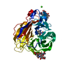
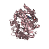
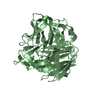
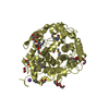
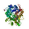
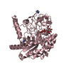
| Title | Crystal structure of cellobiose 2-epimerase in complex with glucosylmannose | |||||||||
 Components Components | Cellobiose 2-epimerase | |||||||||
 Keywords Keywords | ISOMERASE / (alpha/alpha)6 barrel fold / Epimerase / Carbohydrate/Sugar Binding / Epimerization | |||||||||
| Function / homology |  Function and homology information Function and homology informationcellobiose epimerase / cellobiose epimerase activity / carbohydrate metabolic process Similarity search - Function | |||||||||
| Biological species |   Rhodothermus marinus (bacteria) Rhodothermus marinus (bacteria) | |||||||||
| Method |  X-RAY DIFFRACTION / SYNCHROTRON / MOLECULAR REPLACEMENT / Resolution: 1.47 Å X-RAY DIFFRACTION / SYNCHROTRON / MOLECULAR REPLACEMENT / Resolution: 1.47 Å | |||||||||
 Authors Authors | Fujiwara, T. / Saburi, W. / Tanaka, I. / Yao, M. | |||||||||
 Citation Citation | Journal: J.Biol.Chem. / Year: 2014 Title: Structural Insights into the Epimerization of beta-1,4-Linked Oligosaccharides Catalyzed by Cellobiose 2-Epimerase, the Sole Enzyme Epimerizing Non-anomeric Hydroxyl Groups of Unmodified Sugars Authors: Fujiwara, T. / Saburi, W. / Matsui, H. / Mori, H. / Yao, M. | |||||||||
| History |
|
- Structure visualization
| Structure viewer | Molecule: MolmilJmol/JSmol |
|---|
- Downloads & links
-Download
| PDBx/mmCIF format | 3wkg.cif.gz | 113 KB | Display | PDBx/mmCIF format |
|---|---|---|---|---|
| PDB format | pdb3wkg.ent.gz | 82.6 KB | Display | PDB format |
| PDBx/mmJSON format | 3wkg.json.gz | Tree view | PDBx/mmJSON format | |
| Others |  Other downloads Other downloads |
-Validation report
| Arichive directory | https://data.pdbj.org/pub/pdb/validation_reports/wk/3wkgftp://data.pdbj.org/pub/pdb/validation_reports/wk/3wkg | HTTPS FTP |
|---|
-Related structure data
| Related structure data |  3wkfSC  3wkhC  3wkiC S: Starting model for refinement C: citing same article ( |
|---|---|
| Similar structure data |


-Links
 PDBj
PDBj
- Assembly
| Deposited unit | 
| ||||||||
|---|---|---|---|---|---|---|---|---|---|
| 1 |
| ||||||||
| Unit cell |
|
-Components
| #1: Protein | Mass: 47459.355 Da / Num. of mol.: 1 Source method: isolated from a genetically manipulated source Source: (gene. exp.) Rhodothermus marinus (bacteria) / Strain: JCM9785 / Gene: ce / Plasmid: pET22B / Production host: |
|---|---|
| #2: Polysaccharide | beta-D-glucopyranose-(1-4)-beta-D-mannopyranose Source method: isolated from a genetically manipulated source |
| #3: Chemical | ChemComp-CL /   Mass: 35.453 Da / Num. of mol.: 1 / Source method: obtained synthetically / Formula: Cl Mass: 35.453 Da / Num. of mol.: 1 / Source method: obtained synthetically / Formula: Cl |
| #4: Chemical | ChemComp-PO4 /   Mass: 94.971 Da / Num. of mol.: 1 / Source method: obtained synthetically / Formula: PO4 Mass: 94.971 Da / Num. of mol.: 1 / Source method: obtained synthetically / Formula: PO4 |
| #5: Water | ChemComp-HOH /  Mass: 18.015 Da / Num. of mol.: 611 / Source method: isolated from a natural source / Formula: H2O Mass: 18.015 Da / Num. of mol.: 611 / Source method: isolated from a natural source / Formula: H2O |
| Compound details | ONE LIGAND OF THIS STRUCTURE, 4-O-BETA-D-GLUCOSYL-D-MANNOSE(C12H22O11) IS LOCATED AT 502-503, ...ONE LIGAND OF THIS STRUCTURE, 4-O-BETA-D-GLUCOSYL-D-MANNOSE(C12H22O11) IS LOCATED AT 502-503, CHAINE A (REF. LINK RECORD) |
-Experimental details
-Experiment
| Experiment | Method: X-RAY DIFFRACTION / Number of used crystals: 1 |
|---|
- Sample preparation
| Crystal | Density Matthews: 1.82 Å3/Da / Density % sol: 32.52 % |
|---|---|
| Crystal grow | Temperature: 293 K / Method: vapor diffusion, sitting drop / pH: 4.5 Details: 0.1M sodium acetate buffer pH4.5, 1.0M ammonium hydrogen phosphate, VAPOR DIFFUSION, SITTING DROP, temperature 293K |
-Data collection
| Diffraction | Mean temperature: 100 K |
|---|---|
| Diffraction source | Source: SYNCHROTRON / Site: Photon Factory  / Beamline: BL-1A / Wavelength: 1 Å / Beamline: BL-1A / Wavelength: 1 Å |
| Detector | Type: DECTRIS PILATUS 2M-F / Detector: PIXEL / Date: Nov 24, 2012 |
| Radiation | Monochromator: cryocooled channel-cut Si(111) / Protocol: SINGLE WAVELENGTH / Monochromatic (M) / Laue (L): M / Scattering type: x-ray |
| Radiation wavelength | Wavelength: 1 Å / Relative weight: 1 |
| Reflection | Resolution: 1.47→50 Å / Num. obs: 59748 / % possible obs: 99.8 % / Redundancy: 6.4 % / Rmerge(I) obs: 0.076 / Net I/σ(I): 16.8 |
| Reflection shell | Resolution: 1.47→1.56 Å / Redundancy: 6.5 % / Rmerge(I) obs: 0.511 / Mean I/σ(I) obs: 3.8 / Num. unique all: 9460 / % possible all: 99.2 |
- Processing
| Software |
| ||||||||||||||||||||||||
|---|---|---|---|---|---|---|---|---|---|---|---|---|---|---|---|---|---|---|---|---|---|---|---|---|---|
| Refinement | Method to determine structure: MOLECULAR REPLACEMENT Starting model: 3WKF Resolution: 1.47→39.75 Å / SU ML: 0.14 / Isotropic thermal model: isotropic / Cross valid method: THROUGHOUT / σ(F): 1.35 / Phase error: 18 / Stereochemistry target values: ML
| ||||||||||||||||||||||||
| Solvent computation | Shrinkage radii: 0.9 Å / VDW probe radii: 1.11 Å / Solvent model: FLAT BULK SOLVENT MODEL | ||||||||||||||||||||||||
| Refinement step | Cycle: LAST / Resolution: 1.47→39.75 Å
| ||||||||||||||||||||||||
| Refine LS restraints |
| ||||||||||||||||||||||||
| LS refinement shell | Resolution: 1.4704→1.4933 Å
|
