 Movie
Movie Controller

[English] 日本語
 Yorodumi
- PDB-3wjy: Orotidine 5'-monophosphate decarboxylase K72A mutant from M. ther... -
+ Open data

- Basic information
| Entry | Database: PDB / ID: 3wjy | ||||||
|---|---|---|---|---|---|---|---|
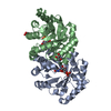
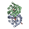
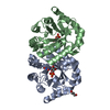
| Title | Orotidine 5'-monophosphate decarboxylase K72A mutant from M. thermoautotrophicus complexed with 6-amino-UMP | ||||||
 Components Components | Orotidine 5'-phosphate decarboxylase | ||||||
 Keywords Keywords | LYASE / protein-ligand complex / TIM barrel / decarboxylase / pyrimidine biosynthesis | ||||||
| Function / homology |  Function and homology information Function and homology informationorotidine-5'-phosphate decarboxylase / orotidine-5'-phosphate decarboxylase activity / 'de novo' UMP biosynthetic process / 'de novo' pyrimidine nucleobase biosynthetic process / cytosol Similarity search - Function | ||||||
| Biological species |   Methanothermobacter thermautotrophicus (archaea) Methanothermobacter thermautotrophicus (archaea) | ||||||
| Method |  X-RAY DIFFRACTION / SYNCHROTRON / MOLECULAR REPLACEMENT / Resolution: 1.72 Å X-RAY DIFFRACTION / SYNCHROTRON / MOLECULAR REPLACEMENT / Resolution: 1.72 Å | ||||||
 Authors Authors | Fujihashi, M. / Pai, E.F. / Miki, K. | ||||||
 Citation Citation | Journal: J.Am.Chem.Soc. / Year: 2013 Title: Substrate distortion contributes to the catalysis of orotidine 5'-monophosphate decarboxylase. Authors: Fujihashi, M. / Ishida, T. / Kuroda, S. / Kotra, L.P. / Pai, E.F. / Miki, K. | ||||||
| History |
|
- Structure visualization
| Structure viewer | Molecule: MolmilJmol/JSmol |
|---|
- Downloads & links
-Download
| PDBx/mmCIF format | 3wjy.cif.gz | 104.4 KB | Display | PDBx/mmCIF format |
|---|---|---|---|---|
| PDB format | pdb3wjy.ent.gz | 79.2 KB | Display | PDB format |
| PDBx/mmJSON format | 3wjy.json.gz | Tree view | PDBx/mmJSON format | |
| Others |  Other downloads Other downloads |
-Validation report
| Arichive directory | https://data.pdbj.org/pub/pdb/validation_reports/wj/3wjyftp://data.pdbj.org/pub/pdb/validation_reports/wj/3wjy | HTTPS FTP |
|---|
-Related structure data
| Related structure data | 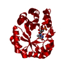 3wjwC 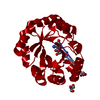 3wjxC  3wjzC 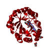 3wk0C 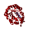 3wk1C 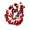 3wk2C 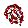 3wk3C  1dvjS C: citing same article ( S: Starting model for refinement |
|---|---|
| Similar structure data |


-Links
 PDBj
PDBj
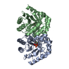
- Assembly
| Deposited unit | 
| ||||||||
|---|---|---|---|---|---|---|---|---|---|
| 1 | 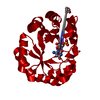
| ||||||||
| Unit cell |
| ||||||||
| Components on special symmetry positions |
|

-Components
| #1: Protein | Mass: 27321.299 Da / Num. of mol.: 1 / Mutation: K72A, L226R, N227I Source method: isolated from a genetically manipulated source Source: (gene. exp.) Methanothermobacter thermautotrophicus (archaea)Strain: ATCC 29096 / DSM 1053 / JCM 10044 / NBRC 100330 / Delta H Gene: pyrF, MTH_129 / Plasmid: pET15b / Production host:  References: UniProt: O26232, orotidine-5'-phosphate decarboxylase |
|---|---|
| #2: Chemical | ChemComp-NUP / 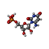  Mass: 339.196 Da / Num. of mol.: 1 / Source method: obtained synthetically / Formula: C9H14N3O9P Mass: 339.196 Da / Num. of mol.: 1 / Source method: obtained synthetically / Formula: C9H14N3O9P |
| #3: Chemical | ChemComp-GOL /   Mass: 92.094 Da / Num. of mol.: 1 / Source method: obtained synthetically / Formula: C3H8O3 Mass: 92.094 Da / Num. of mol.: 1 / Source method: obtained synthetically / Formula: C3H8O3 |
| #4: Water | ChemComp-HOH /  Mass: 18.015 Da / Num. of mol.: 136 / Source method: isolated from a natural source / Formula: H2O Mass: 18.015 Da / Num. of mol.: 136 / Source method: isolated from a natural source / Formula: H2O |
| Sequence details | ACCORDING TO DEPOSITORS, PRO101 IS CORRECT AND UNIPORT IS PROBABLY INCORRECT AT THIS POSITION. ...ACCORDING TO DEPOSITORS |
-Experimental details
-Experiment
| Experiment | Method: X-RAY DIFFRACTION / Number of used crystals: 1 |
|---|
- Sample preparation
| Crystal | Density Matthews: 2.04 Å3/Da / Density % sol: 39.66 % |
|---|---|
| Crystal grow | Temperature: 293 K / Method: vapor diffusion / pH: 6.5 Details: Sodium citrate, pH 6.5, VAPOR DIFFUSION, temperature 293K |
-Data collection
| Diffraction | Mean temperature: 100 K |
|---|---|
| Diffraction source | Source: SYNCHROTRON / Site: APS  / Beamline: 14-BM-C / Wavelength: 0.9 Å / Beamline: 14-BM-C / Wavelength: 0.9 Å |
| Detector | Type: ADSC QUANTUM 315 / Detector: CCD |
| Radiation | Monochromator: Default / Protocol: SINGLE WAVELENGTH / Monochromatic (M) / Laue (L): M / Scattering type: x-ray |
| Radiation wavelength | Wavelength: 0.9 Å / Relative weight: 1 |
| Reflection | Resolution: 1.72→51.78 Å / Num. obs: 24073 / % possible obs: 99.9 % / Rmerge(I) obs: 0.058 / Net I/σ(I): 34.8 |
| Reflection shell | Resolution: 1.72→1.76 Å / Rmerge(I) obs: 0.315 / % possible all: 100 |
- Processing
| Software |
| |||||||||||||||||||||||||||||||||||||||||||||||||||||||||||||||||
|---|---|---|---|---|---|---|---|---|---|---|---|---|---|---|---|---|---|---|---|---|---|---|---|---|---|---|---|---|---|---|---|---|---|---|---|---|---|---|---|---|---|---|---|---|---|---|---|---|---|---|---|---|---|---|---|---|---|---|---|---|---|---|---|---|---|---|
| Refinement | Method to determine structure: MOLECULAR REPLACEMENT Starting model: 1DVJ Resolution: 1.72→50 Å / Cor.coef. Fo:Fc: 0.969 / Cor.coef. Fo:Fc free: 0.96 / Occupancy max: 1 / Occupancy min: 0.3 / SU B: 3.506 / SU ML: 0.053 / Cross valid method: THROUGHOUT / σ(F): 0 / ESU R: 0.092 / ESU R Free: 0.091 / Stereochemistry target values: MAXIMUM LIKELIHOOD
| |||||||||||||||||||||||||||||||||||||||||||||||||||||||||||||||||
| Solvent computation | Ion probe radii: 0.8 Å / Shrinkage radii: 0.8 Å / VDW probe radii: 1.2 Å / Solvent model: MASK | |||||||||||||||||||||||||||||||||||||||||||||||||||||||||||||||||
| Displacement parameters | Biso max: 67.67 Å2 / Biso mean: 19.5217 Å2 / Biso min: 5.58 Å2
| |||||||||||||||||||||||||||||||||||||||||||||||||||||||||||||||||
| Refinement step | Cycle: LAST / Resolution: 1.72→50 Å
| |||||||||||||||||||||||||||||||||||||||||||||||||||||||||||||||||
| Refine LS restraints |
| |||||||||||||||||||||||||||||||||||||||||||||||||||||||||||||||||
| LS refinement shell | Resolution: 1.72→1.765 Å / Total num. of bins used: 20
| |||||||||||||||||||||||||||||||||||||||||||||||||||||||||||||||||
| Refinement TLS params. | Method: refined / Origin x: 18.5892 Å / Origin y: 40.45 Å / Origin z: 29.9529 Å
| |||||||||||||||||||||||||||||||||||||||||||||||||||||||||||||||||
| Refinement TLS group |
|
