 Movie
Movie Controller

[English] 日本語
 Yorodumi
- PDB-3wjk: Crystal structure of Octaprenyl Pyrophosphate synthase from Esche... -
+ Open data

- Basic information
| Entry | Database: PDB / ID: 3wjk | ||||||
|---|---|---|---|---|---|---|---|
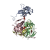
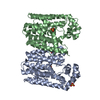
| Title | Crystal structure of Octaprenyl Pyrophosphate synthase from Escherichia coli | ||||||
 Components Components | Octaprenyl diphosphate synthase | ||||||
 Keywords Keywords | TRANSFERASE / prenyltransferase / site-directed mutagenesis / product chain length | ||||||
| Function / homology | Farnesyl Diphosphate Synthase / Farnesyl Diphosphate Synthase / Orthogonal Bundle / Mainly Alpha / :  Function and homology information Function and homology information | ||||||
| Biological species |  | ||||||
| Method |  X-RAY DIFFRACTION / SYNCHROTRON / MOLECULAR REPLACEMENT / Resolution: 2.2 Å X-RAY DIFFRACTION / SYNCHROTRON / MOLECULAR REPLACEMENT / Resolution: 2.2 Å | ||||||
 Authors Authors | Han, X. / Chen, C.C. / Kuo, C.J. / Huang, C.H. / Zheng, Y. / Ko, T.P. / Zhu, Z. / Feng, X. / Oldfield, E. / Liang, P.H. ...Han, X. / Chen, C.C. / Kuo, C.J. / Huang, C.H. / Zheng, Y. / Ko, T.P. / Zhu, Z. / Feng, X. / Oldfield, E. / Liang, P.H. / Guo, R.T. / Ma, Y.H. | ||||||
 Citation Citation | Journal: Proteins / Year: 2015 Title: Crystal structures of ligand-bound octaprenyl pyrophosphate synthase from Escherichia coli reveal the catalytic and chain-length determining mechanisms. Authors: Han, X. / Chen, C.C. / Kuo, C.J. / Huang, C.H. / Zheng, Y. / Ko, T.P. / Zhu, Z. / Feng, X. / Wang, K. / Oldfield, E. / Wang, A.H. / Liang, P.H. / Guo, R.T. / Ma, Y. | ||||||
| History |
|
- Structure visualization
| Structure viewer | Molecule: MolmilJmol/JSmol |
|---|
- Downloads & links
-Download
| PDBx/mmCIF format | 3wjk.cif.gz | 133.3 KB | Display | PDBx/mmCIF format |
|---|---|---|---|---|
| PDB format | pdb3wjk.ent.gz | 103.2 KB | Display | PDB format |
| PDBx/mmJSON format | 3wjk.json.gz | Tree view | PDBx/mmJSON format | |
| Others |  Other downloads Other downloads |
-Validation report
| Arichive directory | https://data.pdbj.org/pub/pdb/validation_reports/wj/3wjkftp://data.pdbj.org/pub/pdb/validation_reports/wj/3wjk | HTTPS FTP |
|---|
-Related structure data
| Related structure data |  3wjnC  3wjoC  3mzvS C: citing same article ( S: Starting model for refinement |
|---|---|
| Similar structure data |


-Links
 PDBj
PDBj- Assembly
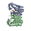
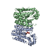
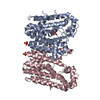
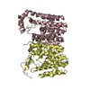
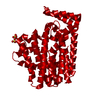
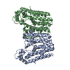
| Deposited unit | 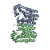
| ||||||||
|---|---|---|---|---|---|---|---|---|---|
| 1 |
| ||||||||
| Unit cell |
|
-Components
| #1: Protein | Mass: 36969.691 Da / Num. of mol.: 2 Source method: isolated from a genetically manipulated source Source: (gene. exp.) References: UniProt: K0BUH0, Transferases; Transferring alkyl or aryl groups, other than methyl groups #2: Water | ChemComp-HOH / |  Mass: 18.015 Da / Num. of mol.: 310 / Source method: isolated from a natural source / Formula: H2O Mass: 18.015 Da / Num. of mol.: 310 / Source method: isolated from a natural source / Formula: H2O |
|---|
-Experimental details
-Experiment
| Experiment | Method: X-RAY DIFFRACTION / Number of used crystals: 1 |
|---|
- Sample preparation
| Crystal | Density Matthews: 2.35 Å3/Da / Density % sol: 47.77 % |
|---|---|
| Crystal grow | Temperature: 298 K / Method: vapor diffusion, sitting drop / pH: 8.5 Details: 0.3M Magnesium chloride hexahydrate, 0.1M Tris-HCl pH 8.5, 24% w/v polyethylene glycol 3350, VAPOR DIFFUSION, SITTING DROP, temperature 298K |
-Data collection
| Diffraction | Mean temperature: 100 K |
|---|---|
| Diffraction source | Source: SYNCHROTRON / Site: NSRRC  / Beamline: BL13B1 / Beamline: BL13B1 |
| Detector | Type: ADSC QUANTUM 315 / Detector: CCD / Date: Aug 26, 2012 |
| Radiation | Protocol: SINGLE WAVELENGTH / Monochromatic (M) / Laue (L): M / Scattering type: x-ray |
| Radiation wavelength | Relative weight: 1 |
| Reflection | Resolution: 2.2→25 Å / Num. obs: 36336 / % possible obs: 99.8 % / Redundancy: 3.9 % / Rmerge(I) obs: 0.058 / Net I/σ(I): 27.5 |
| Reflection shell | Resolution: 2.2→2.28 Å / Redundancy: 3.9 % / Rmerge(I) obs: 0.517 / Mean I/σ(I) obs: 3 / Num. unique all: 13763 / % possible all: 99.4 |
- Processing
| Software |
| |||||||||||||||||||||||||
|---|---|---|---|---|---|---|---|---|---|---|---|---|---|---|---|---|---|---|---|---|---|---|---|---|---|---|
| Refinement | Method to determine structure: MOLECULAR REPLACEMENT Starting model: 3MZV Resolution: 2.2→25 Å / Occupancy max: 1 / Occupancy min: 1 / Stereochemistry target values: Engh & Huber
| |||||||||||||||||||||||||
| Solvent computation | Bsol: 39.727 Å2 | |||||||||||||||||||||||||
| Displacement parameters | Biso max: 90.28 Å2 / Biso mean: 45.9341 Å2 / Biso min: 16.11 Å2
| |||||||||||||||||||||||||
| Refinement step | Cycle: LAST / Resolution: 2.2→25 Å
| |||||||||||||||||||||||||
| Refine LS restraints |
| |||||||||||||||||||||||||
| LS refinement shell | Resolution: 2.2→2.28 Å /
|
