 Movie
Movie Controller

[English] 日本語
 Yorodumi
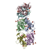
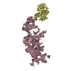
- PDB-3wir: Crystal structure of kojibiose phosphorylase complexed with glucose -
+ Open data

- Basic information
| Entry | Database: PDB / ID: 3wir | ||||||
|---|---|---|---|---|---|---|---|
| Title | Crystal structure of kojibiose phosphorylase complexed with glucose | ||||||
 Components Components | Kojibiose phosphorylase | ||||||
 Keywords Keywords | TRANSFERASE / (alpha/alpha)6 barrel / Phosphorylase | ||||||
| Function / homology |  Function and homology information Function and homology informationkojibiose phosphorylase / kojibiose phosphorylase activity / hydrolase activity, hydrolyzing O-glycosyl compounds / carbohydrate binding / carbohydrate metabolic process Similarity search - Function | ||||||
| Biological species |   Caldicellulosiruptor saccharolyticus (bacteria) Caldicellulosiruptor saccharolyticus (bacteria) | ||||||
| Method |  X-RAY DIFFRACTION / SYNCHROTRON / MOLECULAR REPLACEMENT / Resolution: 2.05 Å X-RAY DIFFRACTION / SYNCHROTRON / MOLECULAR REPLACEMENT / Resolution: 2.05 Å | ||||||
 Authors Authors | Okada, S. / Yamamoto, T. / Watanabe, H. / Nishimoto, T. / Chaen, H. / Fukuda, S. / Wakagi, T. / Fushinobu, S. | ||||||
 Citation Citation | Journal: Febs J. / Year: 2014 Title: Structural and mutational analysis of substrate recognition in kojibiose phosphorylase Authors: Okada, S. / Yamamoto, T. / Watanabe, H. / Nishimoto, T. / Chaen, H. / Fukuda, S. / Wakagi, T. / Fushinobu, S. #1: Journal: Biosci.Biotechnol.Biochem. / Year: 2011 Title: Enzymatic properties of recombinant kojibiose phosphorylase from Caldicellulosiruptor saccharolyticus ATCC43494 Authors: Yamamoto, T. / Nishio-Kosaka, M. / Izawa, S. / Aga, H. / Nishimoto, T. / Chaen, H. / Fukuda, S. | ||||||
| History |
|
- Structure visualization
| Structure viewer | Molecule: MolmilJmol/JSmol |
|---|
- Downloads & links
-Download
| PDBx/mmCIF format | 3wir.cif.gz | 634.2 KB | Display | PDBx/mmCIF format |
|---|---|---|---|---|
| PDB format | pdb3wir.ent.gz | 522.9 KB | Display | PDB format |
| PDBx/mmJSON format | 3wir.json.gz | Tree view | PDBx/mmJSON format | |
| Others |  Other downloads Other downloads |
-Validation report
| Arichive directory | https://data.pdbj.org/pub/pdb/validation_reports/wi/3wirftp://data.pdbj.org/pub/pdb/validation_reports/wi/3wir | HTTPS FTP |
|---|
-Related structure data
| Related structure data | 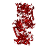 3wiqC 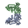 1h54S C: citing same article ( S: Starting model for refinement |
|---|---|
| Similar structure data |


-Links
 PDBj
PDBj


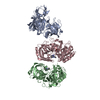
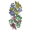
- Assembly
| Deposited unit | 
| ||||||||
|---|---|---|---|---|---|---|---|---|---|
| 1 | 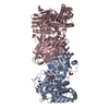
| ||||||||
| 2 | 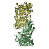
| ||||||||
| Unit cell |
|
-Components
| #1: Protein | Mass: 88685.758 Da / Num. of mol.: 4 Source method: isolated from a genetically manipulated source Source: (gene. exp.) Caldicellulosiruptor saccharolyticus (bacteria)Strain: ATCC 43494 / DSM 8903 / Gene: Csac_0444 / Plasmid: pRSET0439-C-His / Production host: #2: Sugar | ChemComp-BGC /   Type: D-saccharide, beta linking / Mass: 180.156 Da / Num. of mol.: 8 Type: D-saccharide, beta linking / Mass: 180.156 Da / Num. of mol.: 8Source method: isolated from a genetically manipulated source Formula: C6H12O6 #3: Chemical | ChemComp-GOL /   Mass: 92.094 Da / Num. of mol.: 9 / Source method: obtained synthetically / Formula: C3H8O3 Mass: 92.094 Da / Num. of mol.: 9 / Source method: obtained synthetically / Formula: C3H8O3#4: Chemical | ChemComp-PO4 /   Mass: 94.971 Da / Num. of mol.: 6 / Source method: obtained synthetically / Formula: PO4 Mass: 94.971 Da / Num. of mol.: 6 / Source method: obtained synthetically / Formula: PO4#5: Water | ChemComp-HOH / |  Mass: 18.015 Da / Num. of mol.: 1378 / Source method: isolated from a natural source / Formula: H2O Mass: 18.015 Da / Num. of mol.: 1378 / Source method: isolated from a natural source / Formula: H2OHas protein modification | Y | |
|---|
-Experimental details
-Experiment
| Experiment | Method: X-RAY DIFFRACTION / Number of used crystals: 1 |
|---|
- Sample preparation
| Crystal | Density Matthews: 2.43 Å3/Da / Density % sol: 49.46 % |
|---|---|
| Crystal grow | Temperature: 298 K / Method: vapor diffusion, sitting drop / pH: 8.5 Details: 5mM D-glucose, 5mM sodium phosphate, 10%(v/v) 2-propanol, 10%(w/v) PEG3350, 0.1M Tris-HCl, pH 8.5, VAPOR DIFFUSION, SITTING DROP, temperature 298K |
-Data collection
| Diffraction | Mean temperature: 100 K |
|---|---|
| Diffraction source | Source: SYNCHROTRON / Site: SPring-8  / Beamline: BL38B1 / Wavelength: 1 Å / Beamline: BL38B1 / Wavelength: 1 Å |
| Detector | Type: ADSC QUANTUM 315r / Detector: CCD / Date: Jun 24, 2011 |
| Radiation | Monochromator: Fixed exit double crystal monochromator / Protocol: SINGLE WAVELENGTH / Monochromatic (M) / Laue (L): M / Scattering type: x-ray |
| Radiation wavelength | Wavelength: 1 Å / Relative weight: 1 |
| Reflection | Resolution: 2.05→50 Å / Num. all: 209616 / Num. obs: 203954 / % possible obs: 97.3 % / Observed criterion σ(I): -3 / Redundancy: 3.8 % / Rsym value: 0.097 / Net I/σ(I): 14.5 |
| Reflection shell | Resolution: 2.05→2.12 Å / Redundancy: 3.7 % / Mean I/σ(I) obs: 2.3 / Num. unique all: 22589 / Rsym value: 0.46 / % possible all: 96.2 |
- Processing
| Software |
| ||||||||||||||||||||||||||||||||||||||||||||||||||||||||||||||||||||||||||||||||||||||||||||||||||||||||||||||||||||||||||||||||||||||||||||||||||||||||||||||||||||||||||||||||||||||
|---|---|---|---|---|---|---|---|---|---|---|---|---|---|---|---|---|---|---|---|---|---|---|---|---|---|---|---|---|---|---|---|---|---|---|---|---|---|---|---|---|---|---|---|---|---|---|---|---|---|---|---|---|---|---|---|---|---|---|---|---|---|---|---|---|---|---|---|---|---|---|---|---|---|---|---|---|---|---|---|---|---|---|---|---|---|---|---|---|---|---|---|---|---|---|---|---|---|---|---|---|---|---|---|---|---|---|---|---|---|---|---|---|---|---|---|---|---|---|---|---|---|---|---|---|---|---|---|---|---|---|---|---|---|---|---|---|---|---|---|---|---|---|---|---|---|---|---|---|---|---|---|---|---|---|---|---|---|---|---|---|---|---|---|---|---|---|---|---|---|---|---|---|---|---|---|---|---|---|---|---|---|---|---|
| Refinement | Method to determine structure: MOLECULAR REPLACEMENT Starting model: PDB ENTRY 1H54 Resolution: 2.05→46.26 Å / Cor.coef. Fo:Fc: 0.951 / Cor.coef. Fo:Fc free: 0.915 / SU B: 5.21 / SU ML: 0.142 / Cross valid method: THROUGHOUT / ESU R: 0.219 / ESU R Free: 0.194 / Stereochemistry target values: MAXIMUM LIKELIHOOD / Details: HYDROGENS HAVE BEEN ADDED IN THE RIDING POSITIONS
| ||||||||||||||||||||||||||||||||||||||||||||||||||||||||||||||||||||||||||||||||||||||||||||||||||||||||||||||||||||||||||||||||||||||||||||||||||||||||||||||||||||||||||||||||||||||
| Solvent computation | Ion probe radii: 0.8 Å / Shrinkage radii: 0.8 Å / VDW probe radii: 1.2 Å / Solvent model: BABINET MODEL WITH MASK | ||||||||||||||||||||||||||||||||||||||||||||||||||||||||||||||||||||||||||||||||||||||||||||||||||||||||||||||||||||||||||||||||||||||||||||||||||||||||||||||||||||||||||||||||||||||
| Displacement parameters | Biso mean: 31.756 Å2
| ||||||||||||||||||||||||||||||||||||||||||||||||||||||||||||||||||||||||||||||||||||||||||||||||||||||||||||||||||||||||||||||||||||||||||||||||||||||||||||||||||||||||||||||||||||||
| Refinement step | Cycle: LAST / Resolution: 2.05→46.26 Å
| ||||||||||||||||||||||||||||||||||||||||||||||||||||||||||||||||||||||||||||||||||||||||||||||||||||||||||||||||||||||||||||||||||||||||||||||||||||||||||||||||||||||||||||||||||||||
| Refine LS restraints |
|