 Movie
Movie Controller


+ Open data

- Basic information
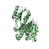
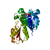
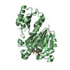
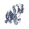
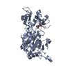
| Entry | Database: PDB / ID: 3wgn | ||||||
|---|---|---|---|---|---|---|---|
| Title | STAPHYLOCOCCUS AUREUS FTSZ bound with GTP-gamma-S | ||||||
 Components Components | Cell division protein FtsZ | ||||||
 Keywords Keywords | CELL CYCLE / FTSZ / GTP-BINDING / TUBULIN HOMOLOG / POLYMERIZATION / GTPASE / CELL DIVISION | ||||||
| Function / homology |  Function and homology information Function and homology informationcell septum / division septum assembly / FtsZ-dependent cytokinesis / cell division site / protein polymerization / GTPase activity / GTP binding / cytoplasm Similarity search - Function | ||||||
| Biological species |   Staphylococcus aureus (bacteria) Staphylococcus aureus (bacteria) | ||||||
| Method |  X-RAY DIFFRACTION / SYNCHROTRON / MOLECULAR REPLACEMENT / molecular replacement / Resolution: 2.606 Å X-RAY DIFFRACTION / SYNCHROTRON / MOLECULAR REPLACEMENT / molecular replacement / Resolution: 2.606 Å | ||||||
 Authors Authors | Matsui, T. / Mogi, N. / Tanaka, I. / Yao, M. | ||||||
 Citation Citation | Journal: J.Biol.Chem. / Year: 2014 Title: Structural change in FtsZ Induced by intermolecular interactions between bound GTP and the T7 loop Authors: Matsui, T. / Han, X. / Yu, J. / Yao, M. / Tanaka, I. | ||||||
| History |
|
- Structure visualization
| Structure viewer | Molecule: MolmilJmol/JSmol |
|---|
- Downloads & links
-Download
| PDBx/mmCIF format | 3wgn.cif.gz | 123.1 KB | Display | PDBx/mmCIF format |
|---|---|---|---|---|
| PDB format | pdb3wgn.ent.gz | 93.8 KB | Display | PDB format |
| PDBx/mmJSON format | 3wgn.json.gz | Tree view | PDBx/mmJSON format | |
| Others |  Other downloads Other downloads |
-Validation report
| Arichive directory | https://data.pdbj.org/pub/pdb/validation_reports/wg/3wgnftp://data.pdbj.org/pub/pdb/validation_reports/wg/3wgn | HTTPS FTP |
|---|
-Related structure data
| Related structure data |  3wgjC  3wgkC  3wglC  3wgmC 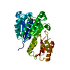 3voaS S: Starting model for refinement C: citing same article ( |
|---|---|
| Similar structure data |


-Links
 PDBj
PDBj


- Assembly
| Deposited unit | 
| ||||||||
|---|---|---|---|---|---|---|---|---|---|
| 1 | 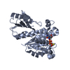
| ||||||||
| 2 | 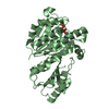
| ||||||||
| Unit cell |
|
-Components
| #1: Protein | Mass: 41274.008 Da / Num. of mol.: 2 Source method: isolated from a genetically manipulated source Source: (gene. exp.) Staphylococcus aureus (bacteria) / Strain: Mu50 / Gene: ftsZ / Plasmid: pET / Production host: #2: Chemical |   Mass: 539.246 Da / Num. of mol.: 2 / Source method: obtained synthetically / Formula: C10H16N5O13P3S Mass: 539.246 Da / Num. of mol.: 2 / Source method: obtained synthetically / Formula: C10H16N5O13P3S |
|---|
-Experimental details
-Experiment
| Experiment | Method: X-RAY DIFFRACTION / Number of used crystals: 1 |
|---|
- Sample preparation
| Crystal | Density Matthews: 1.696 Å3/Da / Density % sol: 27.498 % |
|---|---|
| Crystal grow | Temperature: 293 K / Method: vapor diffusion, sitting drop Details: 1.0M Lithium chloride, 0.1M Sodium acetate, 30%(w/v) PEG6000, 10%(v/v) glycerol, 5mM GTPgammaS, VAPOR DIFFUSION, SITTING DROP, temperature 293K |
-Data collection
| Diffraction | Mean temperature: 100 K | ||||||||||||||||||||||||||||||||||||||||||||||||||||||||||||||||||||||||||||||||||||||||||||||||||||
|---|---|---|---|---|---|---|---|---|---|---|---|---|---|---|---|---|---|---|---|---|---|---|---|---|---|---|---|---|---|---|---|---|---|---|---|---|---|---|---|---|---|---|---|---|---|---|---|---|---|---|---|---|---|---|---|---|---|---|---|---|---|---|---|---|---|---|---|---|---|---|---|---|---|---|---|---|---|---|---|---|---|---|---|---|---|---|---|---|---|---|---|---|---|---|---|---|---|---|---|---|---|
| Diffraction source | Source: SYNCHROTRON / Site: Photon Factory  / Beamline: BL-17A / Wavelength: 0.98 Å / Beamline: BL-17A / Wavelength: 0.98 Å | ||||||||||||||||||||||||||||||||||||||||||||||||||||||||||||||||||||||||||||||||||||||||||||||||||||
| Detector | Type: ADSC QUANTUM 315r / Detector: CCD / Date: Dec 4, 2011 | ||||||||||||||||||||||||||||||||||||||||||||||||||||||||||||||||||||||||||||||||||||||||||||||||||||
| Radiation | Monochromator: Si(111) / Protocol: SINGLE WAVELENGTH / Monochromatic (M) / Laue (L): M / Scattering type: x-ray | ||||||||||||||||||||||||||||||||||||||||||||||||||||||||||||||||||||||||||||||||||||||||||||||||||||
| Radiation wavelength | Wavelength: 0.98 Å / Relative weight: 1 | ||||||||||||||||||||||||||||||||||||||||||||||||||||||||||||||||||||||||||||||||||||||||||||||||||||
| Reflection | Resolution: 2.606→50 Å / Num. obs: 16267 / % possible obs: 98.4 % / Observed criterion σ(I): -3 / Biso Wilson estimate: 53.716 Å2 / Rmerge(I) obs: 0.091 / Net I/σ(I): 11.79 | ||||||||||||||||||||||||||||||||||||||||||||||||||||||||||||||||||||||||||||||||||||||||||||||||||||
| Reflection shell | Diffraction-ID: 1
|
-Phasing
| Phasing | Method: molecular replacement |
|---|
- Processing
| Software |
| |||||||||||||||||||||||||||||||||||||||||||||||||
|---|---|---|---|---|---|---|---|---|---|---|---|---|---|---|---|---|---|---|---|---|---|---|---|---|---|---|---|---|---|---|---|---|---|---|---|---|---|---|---|---|---|---|---|---|---|---|---|---|---|---|
| Refinement | Method to determine structure: MOLECULAR REPLACEMENT Starting model: 3voa Resolution: 2.606→43.511 Å / Occupancy max: 1 / Occupancy min: 1 / FOM work R set: 0.7034 / SU ML: 0.52 / σ(F): 2 / Phase error: 35.46 / Stereochemistry target values: ML
| |||||||||||||||||||||||||||||||||||||||||||||||||
| Solvent computation | Shrinkage radii: 0.9 Å / VDW probe radii: 1.11 Å / Solvent model: FLAT BULK SOLVENT MODEL | |||||||||||||||||||||||||||||||||||||||||||||||||
| Displacement parameters | Biso max: 89.35 Å2 / Biso mean: 38.0887 Å2 / Biso min: 16.81 Å2 | |||||||||||||||||||||||||||||||||||||||||||||||||
| Refinement step | Cycle: LAST / Resolution: 2.606→43.511 Å
| |||||||||||||||||||||||||||||||||||||||||||||||||
| Refine LS restraints |
| |||||||||||||||||||||||||||||||||||||||||||||||||
| LS refinement shell | Refine-ID: X-RAY DIFFRACTION / Total num. of bins used: 6
|
