 Movie
Movie Controller

[English] 日本語
 Yorodumi
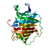
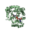
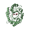
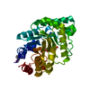
- PDB-3wg6: Crystal structure of conjugated polyketone reductase C1 from Cand... -
+ Open data

- Basic information
| Entry | Database: PDB / ID: 3wg6 | ||||||
|---|---|---|---|---|---|---|---|
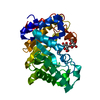
| Title | Crystal structure of conjugated polyketone reductase C1 from Candida parapsilosis complexed with NADPH | ||||||
 Components Components | Conjugated polyketone reductase C1 | ||||||
 Keywords Keywords | OXIDOREDUCTASE / AKR SUPERFAMILY / TIM BARREL / D-PANTOYL LACTONE | ||||||
| Function / homology |  Function and homology information Function and homology information2-dehydropantolactone reductase / 2-dehydropantolactone reductase (A-specific) activity / oxidoreductase activity, acting on NAD(P)H as acceptor / ketone metabolic process / Oxidoreductases; Acting on the CH-OH group of donors; With NAD+ or NADP+ as acceptor Similarity search - Function | ||||||
| Biological species |  Candida parapsilosis (fungus) Candida parapsilosis (fungus) | ||||||
| Method |  X-RAY DIFFRACTION / SYNCHROTRON / MOLECULAR REPLACEMENT / Resolution: 2.2 Å X-RAY DIFFRACTION / SYNCHROTRON / MOLECULAR REPLACEMENT / Resolution: 2.2 Å | ||||||
 Authors Authors | Qin, H.-M. / Yamamura, A. / Miyakawa, T. / Maruoka, S. / Ohtsuka, J. / Nagata, K. / Kataoka, M. / Shimizu, S. / Tanokura, M. | ||||||
 Citation Citation | Journal: Proteins / Year: 2013 Title: Crystal structure of conjugated polyketone reductase (CPR-C1) from Candida parapsilosis IFO 0708 complexed with NADPH. Authors: Qin, H.M. / Yamamura, A. / Miyakawa, T. / Kataoka, M. / Maruoka, S. / Ohtsuka, J. / Nagata, K. / Shimizu, S. / Tanokura, M. | ||||||
| History |
|
- Structure visualization
| Structure viewer | Molecule: MolmilJmol/JSmol |
|---|
- Downloads & links
-Download
| PDBx/mmCIF format | 3wg6.cif.gz | 483.4 KB | Display | PDBx/mmCIF format |
|---|---|---|---|---|
| PDB format | pdb3wg6.ent.gz | 403.9 KB | Display | PDB format |
| PDBx/mmJSON format | 3wg6.json.gz | Tree view | PDBx/mmJSON format | |
| Others |  Other downloads Other downloads |
-Validation report
| Arichive directory | https://data.pdbj.org/pub/pdb/validation_reports/wg/3wg6ftp://data.pdbj.org/pub/pdb/validation_reports/wg/3wg6 | HTTPS FTP |
|---|
-Related structure data
| Similar structure data |
|---|


-Links
 PDBj
PDBj


- Assembly
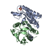
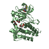
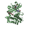
| Deposited unit | 
| ||||||||
|---|---|---|---|---|---|---|---|---|---|
| 1 | 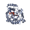
| ||||||||
| 2 | 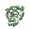
| ||||||||
| 3 | 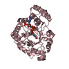
| ||||||||
| 4 | 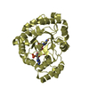
| ||||||||
| Unit cell |
|
-Components
| #1: Protein | Mass: 34821.184 Da / Num. of mol.: 4 Source method: isolated from a genetically manipulated source Source: (gene. exp.) Candida parapsilosis (fungus) / Strain: IFO 0708 / Gene: cpr-c1 / Plasmid: PET-28A / Production host:  #2: Chemical | ChemComp-NDP /   Mass: 745.421 Da / Num. of mol.: 4 / Source method: obtained synthetically / Formula: C21H30N7O17P3 Mass: 745.421 Da / Num. of mol.: 4 / Source method: obtained synthetically / Formula: C21H30N7O17P3#3: Water | ChemComp-HOH / |  Mass: 18.015 Da / Num. of mol.: 326 / Source method: isolated from a natural source / Formula: H2O Mass: 18.015 Da / Num. of mol.: 326 / Source method: isolated from a natural source / Formula: H2O |
|---|
-Experimental details
-Experiment
| Experiment | Method: X-RAY DIFFRACTION / Number of used crystals: 1 |
|---|
- Sample preparation
| Crystal | Density Matthews: 2.44 Å3/Da / Density % sol: 49.55 % |
|---|---|
| Crystal grow | Method: vapor diffusion, sitting drop / pH: 8.5 Details: 5MM NADPH, 0.1M TRIS-HCL, 25% PEG 3350, 5MM NACL, PH 8.5, TEMPERATURE 293K, VAPOR DIFFUSION, SITTING DROP |
-Data collection
| Diffraction | Mean temperature: 100 K |
|---|---|
| Diffraction source | Source: SYNCHROTRON / Site: Photon Factory  / Beamline: AR-NW12A / Wavelength: 1 Å / Beamline: AR-NW12A / Wavelength: 1 Å |
| Detector | Type: ADSC QUANTUM 210 / Detector: CCD / Date: Oct 14, 2009 |
| Radiation | Protocol: SINGLE WAVELENGTH / Monochromatic (M) / Laue (L): M / Scattering type: x-ray |
| Radiation wavelength | Wavelength: 1 Å / Relative weight: 1 |
| Reflection | Resolution: 2.2→20 Å / Num. obs: 67499 / % possible obs: 99.1 % |
- Processing
| Software |
| ||||||||||||||||||||||||||||||||||||||||||||||||||||||||||||||||||||||||||||||||||||||||||||||||||||||||||||||||||||||||||||||||||||||||||||||||||||||||||||||||||||||||||||||||||||||
|---|---|---|---|---|---|---|---|---|---|---|---|---|---|---|---|---|---|---|---|---|---|---|---|---|---|---|---|---|---|---|---|---|---|---|---|---|---|---|---|---|---|---|---|---|---|---|---|---|---|---|---|---|---|---|---|---|---|---|---|---|---|---|---|---|---|---|---|---|---|---|---|---|---|---|---|---|---|---|---|---|---|---|---|---|---|---|---|---|---|---|---|---|---|---|---|---|---|---|---|---|---|---|---|---|---|---|---|---|---|---|---|---|---|---|---|---|---|---|---|---|---|---|---|---|---|---|---|---|---|---|---|---|---|---|---|---|---|---|---|---|---|---|---|---|---|---|---|---|---|---|---|---|---|---|---|---|---|---|---|---|---|---|---|---|---|---|---|---|---|---|---|---|---|---|---|---|---|---|---|---|---|---|---|
| Refinement | Method to determine structure: MOLECULAR REPLACEMENT / Resolution: 2.2→19.78 Å / Cor.coef. Fo:Fc: 0.952 / Cor.coef. Fo:Fc free: 0.919 / SU B: 13.719 / SU ML: 0.156 / Cross valid method: THROUGHOUT / σ(F): 0 / ESU R Free: 0.216 / Stereochemistry target values: MAXIMUM LIKELIHOOD / Details: HYDROGENS HAVE BEEN ADDED IN THE RIDING POSITIONS
| ||||||||||||||||||||||||||||||||||||||||||||||||||||||||||||||||||||||||||||||||||||||||||||||||||||||||||||||||||||||||||||||||||||||||||||||||||||||||||||||||||||||||||||||||||||||
| Solvent computation | Ion probe radii: 0.8 Å / Shrinkage radii: 0.8 Å / VDW probe radii: 1.4 Å / Solvent model: MASK | ||||||||||||||||||||||||||||||||||||||||||||||||||||||||||||||||||||||||||||||||||||||||||||||||||||||||||||||||||||||||||||||||||||||||||||||||||||||||||||||||||||||||||||||||||||||
| Displacement parameters | Biso mean: 32.3 Å2
| ||||||||||||||||||||||||||||||||||||||||||||||||||||||||||||||||||||||||||||||||||||||||||||||||||||||||||||||||||||||||||||||||||||||||||||||||||||||||||||||||||||||||||||||||||||||
| Refinement step | Cycle: LAST / Resolution: 2.2→19.78 Å
| ||||||||||||||||||||||||||||||||||||||||||||||||||||||||||||||||||||||||||||||||||||||||||||||||||||||||||||||||||||||||||||||||||||||||||||||||||||||||||||||||||||||||||||||||||||||
| Refine LS restraints |
|