 Movie
Movie Controller


+ Open data

- Basic information
| Entry | Database: PDB / ID: 3w7t | |||||||||
|---|---|---|---|---|---|---|---|---|---|---|
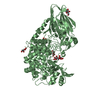
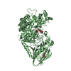
| Title | Escherichia coli K12 YgjK complexed with mannose | |||||||||
 Components Components | Uncharacterized protein YgjK | |||||||||
 Keywords Keywords | HYDROLASE / GH63 / processing alpha-glucosidase I / alpha/alpha barrel | |||||||||
| Function / homology |  Function and homology information Function and homology informationglucosidase complex / alpha,alpha-trehalase activity / trehalose catabolic process / glucosidase activity / Hydrolases; Glycosylases; Glycosidases, i.e. enzymes that hydrolyse O- and S-glycosyl compounds / oligosaccharide catabolic process / DNA damage response / metal ion binding Similarity search - Function | |||||||||
| Biological species |  | |||||||||
| Method |  X-RAY DIFFRACTION / SYNCHROTRON / MOLECULAR REPLACEMENT / Resolution: 1.5 Å X-RAY DIFFRACTION / SYNCHROTRON / MOLECULAR REPLACEMENT / Resolution: 1.5 Å | |||||||||
 Authors Authors | Miyazaki, T. / Kurakata, Y. / Uechi, A. / Yoshida, H. / Kamitori, S. / Sakano, Y. / Nishikawa, A. / Tonozuka, T. | |||||||||
 Citation Citation | Journal: J.Mol.Biol. / Year: 2008 Title: Structural insights into the substrate specificity and function of Escherichia coli K12 YgjK, a glucosidase belonging to the glycoside hydrolase family 63. Authors: Kurakata, Y. / Uechi, A. / Yoshida, H. / Kamitori, S. / Sakano, Y. / Nishikawa, A. / Tonozuka, T. | |||||||||
| History |
|
- Structure visualization
| Structure viewer | Molecule: MolmilJmol/JSmol |
|---|
- Downloads & links
-Download
| PDBx/mmCIF format | 3w7t.cif.gz | 347.9 KB | Display | PDBx/mmCIF format |
|---|---|---|---|---|
| PDB format | pdb3w7t.ent.gz | 281 KB | Display | PDB format |
| PDBx/mmJSON format | 3w7t.json.gz | Tree view | PDBx/mmJSON format | |
| Others |  Other downloads Other downloads |
-Validation report
| Arichive directory | https://data.pdbj.org/pub/pdb/validation_reports/w7/3w7tftp://data.pdbj.org/pub/pdb/validation_reports/w7/3w7t | HTTPS FTP |
|---|
-Related structure data
| Related structure data |  3d3iC  3w7sC  3w7uC  2ds3 C: citing same article ( S: Starting model for refinement |
|---|---|
| Similar structure data |


-Links
 PDBj
PDBj
- Assembly
| Deposited unit | 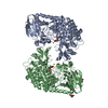
| ||||||||
|---|---|---|---|---|---|---|---|---|---|
| 1 | 
| ||||||||
| 2 | 
| ||||||||
| Unit cell |
|
-Components
| #1: Protein | Mass: 86004.820 Da / Num. of mol.: 2 Source method: isolated from a genetically manipulated source Source: (gene. exp.) #2: Chemical |   Mass: 40.078 Da / Num. of mol.: 2 / Source method: obtained synthetically / Formula: Ca Mass: 40.078 Da / Num. of mol.: 2 / Source method: obtained synthetically / Formula: Ca#3: Chemical |   Mass: 24.305 Da / Num. of mol.: 2 / Source method: obtained synthetically / Formula: Mg Mass: 24.305 Da / Num. of mol.: 2 / Source method: obtained synthetically / Formula: Mg#4: Sugar | ChemComp-BMA /   Type: D-saccharide, beta linking / Mass: 180.156 Da / Num. of mol.: 7 Type: D-saccharide, beta linking / Mass: 180.156 Da / Num. of mol.: 7Source method: isolated from a genetically manipulated source Formula: C6H12O6 #5: Water | ChemComp-HOH / |  Mass: 18.015 Da / Num. of mol.: 1656 / Source method: isolated from a natural source / Formula: H2O Mass: 18.015 Da / Num. of mol.: 1656 / Source method: isolated from a natural source / Formula: H2OHas protein modification | Y | |
|---|
-Experimental details
-Experiment
| Experiment | Method: X-RAY DIFFRACTION / Number of used crystals: 1 |
|---|
- Sample preparation
| Crystal | Density Matthews: 2.14 Å3/Da / Density % sol: 42.64 % |
|---|---|
| Crystal grow | Temperature: 293 K / Method: vapor diffusion, hanging drop / pH: 5.8 Details: 20% PEG 8000, 0.6M magnesium chloride, 100mM Tris-HCl buffer, pH 5.8, VAPOR DIFFUSION, HANGING DROP, temperature 293K, temperature 293.0K |
-Data collection
| Diffraction | Mean temperature: 100 K |
|---|---|
| Diffraction source | Source: SYNCHROTRON / Site: Photon Factory  / Beamline: BL-17A / Wavelength: 1 Å / Beamline: BL-17A / Wavelength: 1 Å |
| Detector | Type: ADSC QUANTUM 4 / Detector: CCD / Date: May 20, 2007 |
| Radiation | Protocol: SINGLE WAVELENGTH / Monochromatic (M) / Laue (L): M / Scattering type: x-ray |
| Radiation wavelength | Wavelength: 1 Å / Relative weight: 1 |
| Reflection | Resolution: 1.5→50 Å / Num. obs: 222770 / % possible obs: 96.6 % / Rmerge(I) obs: 0.063 |
| Reflection shell | Resolution: 1.5→1.55 Å / Rmerge(I) obs: 0.388 / Mean I/σ(I) obs: 3.93 / % possible all: 98.8 |
- Processing
| Software |
| ||||||||||||||||||||||||||||||||||||||||||||||||||||||||||||||||||||||||||||||||||||||||||||||||||||||||||||||||||||||||||||||||||||||||||||||||||||||||||||||||||||||||||
|---|---|---|---|---|---|---|---|---|---|---|---|---|---|---|---|---|---|---|---|---|---|---|---|---|---|---|---|---|---|---|---|---|---|---|---|---|---|---|---|---|---|---|---|---|---|---|---|---|---|---|---|---|---|---|---|---|---|---|---|---|---|---|---|---|---|---|---|---|---|---|---|---|---|---|---|---|---|---|---|---|---|---|---|---|---|---|---|---|---|---|---|---|---|---|---|---|---|---|---|---|---|---|---|---|---|---|---|---|---|---|---|---|---|---|---|---|---|---|---|---|---|---|---|---|---|---|---|---|---|---|---|---|---|---|---|---|---|---|---|---|---|---|---|---|---|---|---|---|---|---|---|---|---|---|---|---|---|---|---|---|---|---|---|---|---|---|---|---|---|---|---|
| Refinement | Method to determine structure: MOLECULAR REPLACEMENT Starting model: 2DS3 2ds3 Resolution: 1.5→46.03 Å / Cor.coef. Fo:Fc: 0.972 / Cor.coef. Fo:Fc free: 0.962 / SU B: 1.185 / SU ML: 0.045 / Cross valid method: THROUGHOUT / ESU R: 0.076 / ESU R Free: 0.075 / Stereochemistry target values: MAXIMUM LIKELIHOOD / Details: HYDROGENS HAVE BEEN ADDED IN THE RIDING POSITIONS
| ||||||||||||||||||||||||||||||||||||||||||||||||||||||||||||||||||||||||||||||||||||||||||||||||||||||||||||||||||||||||||||||||||||||||||||||||||||||||||||||||||||||||||
| Solvent computation | Ion probe radii: 0.8 Å / Shrinkage radii: 0.8 Å / VDW probe radii: 1.2 Å / Solvent model: BABINET MODEL WITH MASK | ||||||||||||||||||||||||||||||||||||||||||||||||||||||||||||||||||||||||||||||||||||||||||||||||||||||||||||||||||||||||||||||||||||||||||||||||||||||||||||||||||||||||||
| Displacement parameters | Biso mean: 15.792 Å2
| ||||||||||||||||||||||||||||||||||||||||||||||||||||||||||||||||||||||||||||||||||||||||||||||||||||||||||||||||||||||||||||||||||||||||||||||||||||||||||||||||||||||||||
| Refinement step | Cycle: LAST / Resolution: 1.5→46.03 Å
| ||||||||||||||||||||||||||||||||||||||||||||||||||||||||||||||||||||||||||||||||||||||||||||||||||||||||||||||||||||||||||||||||||||||||||||||||||||||||||||||||||||||||||
| Refine LS restraints |
| ||||||||||||||||||||||||||||||||||||||||||||||||||||||||||||||||||||||||||||||||||||||||||||||||||||||||||||||||||||||||||||||||||||||||||||||||||||||||||||||||||||||||||
| LS refinement shell | Resolution: 1.5→1.539 Å / Total num. of bins used: 20
|
