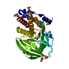
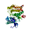
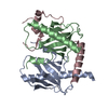
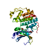
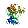
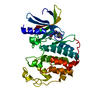
Entry Database : PDB / ID : 3w33Title EGFR kinase domain complexed with compound 19b Epidermal growth factor receptor Keywords / / / / / / Function / homology Function Domain/homology Component
/ / / / / / / / / / / / / / / / / / / / / / / / / / / / / / / / / / / / / / / / / / / / / / / / / / / / / / / / / / / / / / / / / / / / / / / / / / / / / / / / / / / / / / / / / / / / / / / / / / / / / / / / / / / / / / / / / / / / / / / / / / / / / / / / / / / / / / / / / / / / / / / / / / / / / Biological species Homo sapiens (human)Method / / / Resolution : 1.7 Å Authors Sogabe, S. / Kawakita, Y. Journal : Bioorg.Med.Chem. / Year : 2013Title : Design and synthesis of novel pyrimido[4,5-b]azepine derivatives as HER2/EGFR dual inhibitorsAuthors : Kawakita, Y. / Seto, M. / Ohashi, T. / Tamura, T. / Yusa, T. / Miki, H. / Iwata, H. / Kamiguchi, H. / Tanaka, T. / Sogabe, S. / Ohta, Y. / Ishikawa, T. History Deposition Dec 7, 2012 Deposition site / Processing site Revision 1.0 Mar 6, 2013 Provider / Type Revision 1.1 Aug 14, 2013 Group Revision 1.2 Nov 8, 2023 Group Data collection / Database references ... Data collection / Database references / Derived calculations / Refinement description Category chem_comp_atom / chem_comp_bond ... chem_comp_atom / chem_comp_bond / database_2 / pdbx_initial_refinement_model / struct_ref_seq_dif / struct_site Item _database_2.pdbx_DOI / _database_2.pdbx_database_accession ... _database_2.pdbx_DOI / _database_2.pdbx_database_accession / _struct_ref_seq_dif.details / _struct_site.pdbx_auth_asym_id / _struct_site.pdbx_auth_comp_id / _struct_site.pdbx_auth_seq_id
Show all Show less
 Movie
Movie Controller


 Open data
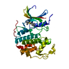
 Basic information
 Components
 Keywords
 Function and homology information
 Homo sapiens (human)
 X-RAY DIFFRACTION /
  Authors
 Citation
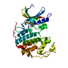
 Structure visualization
 Downloads & links
 Other downloads


 PDBj
PDBj


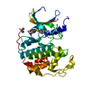
 Assembly
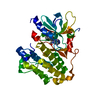

 Spodoptera frugiperda (fall armyworm)
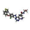
 Mass: 507.992 Da / Num. of mol.: 1 / Source method: obtained synthetically / Formula: C25H22ClN5O3S
 Mass: 96.063 Da / Num. of mol.: 1 / Source method: obtained synthetically / Formula: SO4
 Mass: 18.015 Da / Num. of mol.: 127 / Source method: isolated from a natural source / Formula: H2O
 Sample preparation
 / Beamline: 5.0.3 / Wavelength: 0.97649 Å
 Processing