 Movie
Movie Controller

[English] 日本語
 Yorodumi
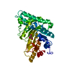
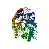
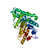
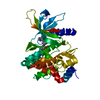
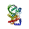
- PDB-3w24: The high-resolution crystal structure of TsXylA, intracellular xy... -
+ Open data

- Basic information
| Entry | Database: PDB / ID: 3w24 | ||||||
|---|---|---|---|---|---|---|---|
| Title | The high-resolution crystal structure of TsXylA, intracellular xylanase from Thermoanaerobacterium saccharolyticum JW/SL-YS485 | ||||||
 Components Components | Glycoside hydrolase family 10 | ||||||
 Keywords Keywords | HYDROLASE / glycoside hydrolase / xylanase / thermophilic | ||||||
| Function / homology |  Function and homology information Function and homology informationendo-1,4-beta-xylanase / endo-1,4-beta-xylanase activity / xylan catabolic process / carbohydrate binding Similarity search - Function | ||||||
| Biological species |  Thermoanaerobacterium saccharolyticum (bacteria) Thermoanaerobacterium saccharolyticum (bacteria) | ||||||
| Method |  X-RAY DIFFRACTION / SYNCHROTRON / MOLECULAR REPLACEMENT / Resolution: 1.35 Å X-RAY DIFFRACTION / SYNCHROTRON / MOLECULAR REPLACEMENT / Resolution: 1.35 Å | ||||||
 Authors Authors | Han, X. / Gao, J. / Shang, N. / Huang, C.-H. / Ko, T.-P. / Zhu, Z. / Wiegel, J. / Shao, W. / Guo, R.-T. | ||||||
 Citation Citation | Journal: Proteins / Year: 2013 Title: Structural and functional analyses of catalytic domain of GH10 xylanase from Thermoanaerobacterium saccharolyticum JW/SL-YS485 Authors: Han, X. / Gao, J. / Shang, N. / Huang, C.-H. / Ko, T.-P. / Chen, C.C. / Chan, H.C. / Cheng, Y.S. / Zhu, Z. / Wiegel, J. / Luo, W. / Guo, R.-T. / Ma, Y. | ||||||
| History |
|
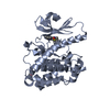
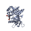
- Structure visualization
| Structure viewer | Molecule: MolmilJmol/JSmol |
|---|
- Downloads & links
-Download
| PDBx/mmCIF format | 3w24.cif.gz | 92.4 KB | Display | PDBx/mmCIF format |
|---|---|---|---|---|
| PDB format | pdb3w24.ent.gz | 68.2 KB | Display | PDB format |
| PDBx/mmJSON format | 3w24.json.gz | Tree view | PDBx/mmJSON format | |
| Others |  Other downloads Other downloads |
-Validation report
| Arichive directory | https://data.pdbj.org/pub/pdb/validation_reports/w2/3w24ftp://data.pdbj.org/pub/pdb/validation_reports/w2/3w24 | HTTPS FTP |
|---|
-Related structure data
| Related structure data |  3w25C  3w26C  3w27C  3w28C  3w29C  2q8xS  3w2b C: citing same article ( S: Starting model for refinement |
|---|---|
| Similar structure data |


-Links
 PDBj
PDBj
- Assembly
| Deposited unit | 
| |||||||||||||||
|---|---|---|---|---|---|---|---|---|---|---|---|---|---|---|---|---|
| 1 |
| |||||||||||||||
| Unit cell |
| |||||||||||||||
| Components on special symmetry positions |
|
-Components
| #1: Protein | Mass: 38063.055 Da / Num. of mol.: 1 / Fragment: UNP residues 349-684 / Mutation: P326L Source method: isolated from a genetically manipulated source Source: (gene. exp.) Thermoanaerobacterium saccharolyticum (bacteria)Strain: JW/SL-YS485 / Gene: Tsac_1459 / Plasmid: pET46a / Production host: |
|---|---|
| #2: Water | ChemComp-HOH /  Mass: 18.015 Da / Num. of mol.: 605 / Source method: isolated from a natural source / Formula: H2O Mass: 18.015 Da / Num. of mol.: 605 / Source method: isolated from a natural source / Formula: H2O |
-Experimental details
-Experiment
| Experiment | Method: X-RAY DIFFRACTION / Number of used crystals: 1 |
|---|
- Sample preparation
| Crystal | Density Matthews: 2.67 Å3/Da / Density % sol: 54 % |
|---|---|
| Crystal grow | Temperature: 295 K / Method: vapor diffusion, sitting drop / pH: 8 Details: 0.1M Tris, 25% PEG 6000, 0.8M LiCl, pH 8.0, VAPOR DIFFUSION, SITTING DROP, temperature 295K |
-Data collection
| Diffraction | Mean temperature: 100 K |
|---|---|
| Diffraction source | Source: SYNCHROTRON / Site: NSRRC  / Beamline: BL13B1 / Beamline: BL13B1 |
| Detector | Type: ADSC QUANTUM 315 / Detector: CCD / Date: Jun 6, 2011 |
| Radiation | Protocol: SINGLE WAVELENGTH / Monochromatic (M) / Laue (L): M / Scattering type: x-ray |
| Radiation wavelength | Relative weight: 1 |
| Reflection | Resolution: 1.35→25 Å / Num. obs: 90031 / % possible obs: 97.6 % / Redundancy: 3.6 % / Rmerge(I) obs: 0.062 / Net I/σ(I): 21.8 |
| Reflection shell | Resolution: 1.35→1.4 Å / Redundancy: 3.6 % / Rmerge(I) obs: 0.481 / Mean I/σ(I) obs: 3.8 / % possible all: 100 |
- Processing
| Software |
| ||||||||||||||||||||
|---|---|---|---|---|---|---|---|---|---|---|---|---|---|---|---|---|---|---|---|---|---|
| Refinement | Method to determine structure: MOLECULAR REPLACEMENT Starting model: 2Q8X Resolution: 1.35→23.63 Å / Stereochemistry target values: Engh & Huber
| ||||||||||||||||||||
| Solvent computation | Bsol: 53.6594 Å2 | ||||||||||||||||||||
| Displacement parameters | Biso mean: 15.9421 Å2
| ||||||||||||||||||||
| Refinement step | Cycle: LAST / Resolution: 1.35→23.63 Å
| ||||||||||||||||||||
| Refine LS restraints |
|
