 Movie
Movie Controller

[English] 日本語
 Yorodumi
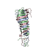
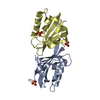
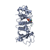
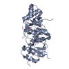
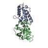
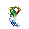
- PDB-3vtn: The crystal structure of the C-terminal domain of Mu phage centra... -
+ Open data

- Basic information
| Entry | Database: PDB / ID: 3vtn | ||||||
|---|---|---|---|---|---|---|---|
| Title | The crystal structure of the C-terminal domain of Mu phage central spike - Pt derivative for MAD | ||||||
 Components Components | Protein gp45 | ||||||
 Keywords Keywords | METAL BINDING PROTEIN / beta-helix / central spike / Mu phage | ||||||
| Function / homology |  Function and homology information Function and homology informationsymbiont genome ejection through host cell envelope, contractile tail mechanism / virus tail, baseplate / viral tail assembly / host cell cytoplasm / metal ion binding Similarity search - Function | ||||||
| Biological species |  Enterobacteria phage Mu (virus) Enterobacteria phage Mu (virus) | ||||||
| Method |  X-RAY DIFFRACTION / SYNCHROTRON / MAD / Resolution: 1.75 Å X-RAY DIFFRACTION / SYNCHROTRON / MAD / Resolution: 1.75 Å | ||||||
 Authors Authors | Harada, K. / Yamashita, E. / Nakagawa, A. / Takeda, S. | ||||||
 Citation Citation | Journal: Biochim.Biophys.Acta / Year: 2013 Title: Crystal structure of the C-terminal domain of Mu phage central spike and functions of bound calcium ion Authors: Harada, K. / Yamashita, E. / Nakagawa, A. / Miyafusa, T. / Tsumoto, K. / Ueno, T. / Toyama, Y. / Takeda, S. | ||||||
| History |
|
- Structure visualization
| Structure viewer | Molecule: MolmilJmol/JSmol |
|---|
- Downloads & links
-Download
| PDBx/mmCIF format | 3vtn.cif.gz | 26.9 KB | Display | PDBx/mmCIF format |
|---|---|---|---|---|
| PDB format | pdb3vtn.ent.gz | 16.7 KB | Display | PDB format |
| PDBx/mmJSON format | 3vtn.json.gz | Tree view | PDBx/mmJSON format | |
| Others |  Other downloads Other downloads |
-Validation report
| Arichive directory | https://data.pdbj.org/pub/pdb/validation_reports/vt/3vtnftp://data.pdbj.org/pub/pdb/validation_reports/vt/3vtn | HTTPS FTP |
|---|
-Related structure data


-Links
 PDBj
PDBj
- Assembly
| Deposited unit | 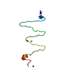
| ||||||||
|---|---|---|---|---|---|---|---|---|---|
| 1 | 
| ||||||||
| Unit cell |
|
-Components
| #1: Protein | Mass: 10912.162 Da / Num. of mol.: 1 / Fragment: UNP residues 100-197 Source method: isolated from a genetically manipulated source Source: (gene. exp.) Enterobacteria phage Mu (virus) / Gene: 45 / Production host:  |
|---|---|
| #2: Chemical | ChemComp-PT /   Mass: 195.078 Da / Num. of mol.: 1 / Source method: obtained synthetically / Formula: Pt Mass: 195.078 Da / Num. of mol.: 1 / Source method: obtained synthetically / Formula: Pt |
| #3: Chemical | ChemComp-FE /   Mass: 55.845 Da / Num. of mol.: 1 / Source method: obtained synthetically / Formula: Fe Mass: 55.845 Da / Num. of mol.: 1 / Source method: obtained synthetically / Formula: Fe |
| #4: Water | ChemComp-HOH /  Mass: 18.015 Da / Num. of mol.: 11 / Source method: isolated from a natural source / Formula: H2O Mass: 18.015 Da / Num. of mol.: 11 / Source method: isolated from a natural source / Formula: H2O |
-Experimental details
-Experiment
| Experiment | Method: X-RAY DIFFRACTION / Number of used crystals: 1 |
|---|
- Sample preparation
| Crystal | Density Matthews: 2.12 Å3/Da / Density % sol: 42.05 % |
|---|---|
| Crystal grow | Temperature: 293 K / Method: vapor diffusion, hanging drop / pH: 4.5 Details: 21% PEG 8000, 0.1M acetate pH 4.5, 0.2M sodium chloride, VAPOR DIFFUSION, HANGING DROP, temperature 293K |
-Data collection
| Diffraction | Mean temperature: 100 K | ||||||||||||
|---|---|---|---|---|---|---|---|---|---|---|---|---|---|
| Diffraction source | Source: SYNCHROTRON / Site: SPring-8  / Beamline: BL44XU / Wavelength: 1.0717, 1.0721, 1.0539 / Beamline: BL44XU / Wavelength: 1.0717, 1.0721, 1.0539 | ||||||||||||
| Detector | Type: RAYONIX MX225HE / Detector: CCD / Date: Sep 30, 2011 | ||||||||||||
| Radiation | Protocol: MAD / Monochromatic (M) / Laue (L): M / Scattering type: x-ray | ||||||||||||
| Radiation wavelength |
| ||||||||||||
| Reflection | Resolution: 1.75→36.06 Å / Num. obs: 9825 |
- Processing
| Software |
| ||||||||||||||||||||||||||||||||||||||||||||||||||||||||||||||||||||||||||||||||||||||||||||||||||||||||||||||||||||||||||||||||||||||||||||||||||||||||||||||||||||||||||
|---|---|---|---|---|---|---|---|---|---|---|---|---|---|---|---|---|---|---|---|---|---|---|---|---|---|---|---|---|---|---|---|---|---|---|---|---|---|---|---|---|---|---|---|---|---|---|---|---|---|---|---|---|---|---|---|---|---|---|---|---|---|---|---|---|---|---|---|---|---|---|---|---|---|---|---|---|---|---|---|---|---|---|---|---|---|---|---|---|---|---|---|---|---|---|---|---|---|---|---|---|---|---|---|---|---|---|---|---|---|---|---|---|---|---|---|---|---|---|---|---|---|---|---|---|---|---|---|---|---|---|---|---|---|---|---|---|---|---|---|---|---|---|---|---|---|---|---|---|---|---|---|---|---|---|---|---|---|---|---|---|---|---|---|---|---|---|---|---|---|---|---|
| Refinement | Method to determine structure: MAD / Resolution: 1.75→36.06 Å / Cor.coef. Fo:Fc: 0.913 / Cor.coef. Fo:Fc free: 0.875 / SU B: 2.701 / SU ML: 0.089 / Cross valid method: THROUGHOUT / ESU R: 0.137 / ESU R Free: 0.133 / Stereochemistry target values: MAXIMUM LIKELIHOOD / Details: HYDROGENS HAVE BEEN ADDED IN THE RIDING POSITIONS
| ||||||||||||||||||||||||||||||||||||||||||||||||||||||||||||||||||||||||||||||||||||||||||||||||||||||||||||||||||||||||||||||||||||||||||||||||||||||||||||||||||||||||||
| Solvent computation | Ion probe radii: 0.8 Å / Shrinkage radii: 0.8 Å / VDW probe radii: 1.4 Å / Solvent model: MASK | ||||||||||||||||||||||||||||||||||||||||||||||||||||||||||||||||||||||||||||||||||||||||||||||||||||||||||||||||||||||||||||||||||||||||||||||||||||||||||||||||||||||||||
| Displacement parameters | Biso mean: 25.963 Å2
| ||||||||||||||||||||||||||||||||||||||||||||||||||||||||||||||||||||||||||||||||||||||||||||||||||||||||||||||||||||||||||||||||||||||||||||||||||||||||||||||||||||||||||
| Refinement step | Cycle: LAST / Resolution: 1.75→36.06 Å
| ||||||||||||||||||||||||||||||||||||||||||||||||||||||||||||||||||||||||||||||||||||||||||||||||||||||||||||||||||||||||||||||||||||||||||||||||||||||||||||||||||||||||||
| Refine LS restraints |
| ||||||||||||||||||||||||||||||||||||||||||||||||||||||||||||||||||||||||||||||||||||||||||||||||||||||||||||||||||||||||||||||||||||||||||||||||||||||||||||||||||||||||||
| LS refinement shell | Resolution: 1.75→1.795 Å / Total num. of bins used: 20
|
