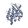
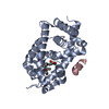
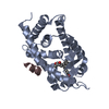
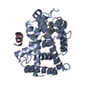
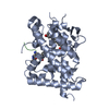
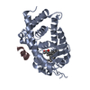
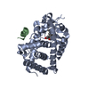
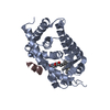
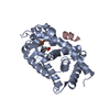
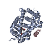
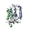
Entry Database : PDB / ID : 3vrtTitle VDR ligand binding domain in complex with 2-Mehylidene-19,25,26,27-tetranor-1alpha,24-dihydroxyvitaminD3 13-meric peptide from Mediator of RNA polymerase II transcription subunit 1 Vitamin D3 receptor Keywords / / / / / Function / homology Function Domain/homology Component
/ / / / / / / / / / / / / / / / / / / / / / / / / / / / / / / / / / / / / / / / / / / / / / / / / / / / / / / / / / / / / / / / / / / / / / / / / / / / / / / / / / / / / / / / / / / / / / / / / / / / / / / / / / / / / / / / / / / / / / / / / / / / / / / / / / / / / / / / / / Biological species Rattus norvegicus (Norway rat)Homo sapiens (human)Method / / / Resolution : 2.4 Å Authors Nakabayashi, M. / Yoshimoto, N. / Inaba, Y. / Itoh, T. / Ito, N. / Yamamoto, K. Journal : J.Med.Chem. / Year : 2012Title : Butyl pocket formation in the vitamin d receptor strongly affects the agonistic or antagonistic behavior of ligandsAuthors : Yoshimoto, N. / Sakamaki, Y. / Haeta, M. / Kato, A. / Inaba, Y. / Itoh, T. / Nakabayashi, M. / Ito, N. / Yamamoto, K. History Deposition Apr 14, 2012 Deposition site / Processing site Revision 1.0 May 23, 2012 Provider / Type Revision 1.1 Mar 20, 2024 Group / Database references / Derived calculationsCategory chem_comp_atom / chem_comp_bond ... chem_comp_atom / chem_comp_bond / database_2 / struct_ref_seq_dif / struct_site Item _database_2.pdbx_DOI / _database_2.pdbx_database_accession ... _database_2.pdbx_DOI / _database_2.pdbx_database_accession / _struct_ref_seq_dif.details / _struct_site.pdbx_auth_asym_id / _struct_site.pdbx_auth_comp_id / _struct_site.pdbx_auth_seq_id
Show all Show less
 Movie
Movie Controller

 Yorodumi
 Open data
 Basic information
 Components
 Keywords
 Function and homology information
 Homo sapiens (human)
 X-RAY DIFFRACTION /
  Authors
 Citation
 Structure visualization
 Downloads & links
 Other downloads
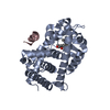

 PDBj
PDBj


 Assembly


 Mass: 374.557 Da / Num. of mol.: 1 / Source method: obtained synthetically / Formula: C24H38O3
 Mass: 18.015 Da / Num. of mol.: 40 / Source method: isolated from a natural source / Formula: H2O
 Sample preparation
 / Beamline: BL-6A / Wavelength: 0.978 Å
 Processing