 Movie
Movie Controller

[English] 日本語
 Yorodumi
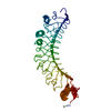
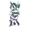
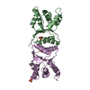
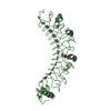
- PDB-3vnc: Crystal Structure of TIP-alpha N25 from Helicobacter Pylori in it... -
+ Open data

- Basic information
| Entry | Database: PDB / ID: 3vnc | ||||||
|---|---|---|---|---|---|---|---|
| Title | Crystal Structure of TIP-alpha N25 from Helicobacter Pylori in its natural dimeric form | ||||||
 Components Components | TIP-alpha | ||||||
 Keywords Keywords | DNA BINDING PROTEIN / TNF-alpha-inducing Protein / HP0596 / Homodimer / Carcinogenic Factor | ||||||
| Function / homology | Helicobacter TNF-alpha-Inducing protein / Helicobacter TNF-alpha-inducing protein / TNF-alpha-Inducing protein of Helicobacter / Thiol Ester Dehydrase; Chain A / Prokaryotic membrane lipoprotein lipid attachment site profile. / Roll / Alpha Beta / Tumor necrosis factor alpha-inducing protein Function and homology information Function and homology information | ||||||
| Biological species |   Helicobacter pylori (bacteria) Helicobacter pylori (bacteria) | ||||||
| Method |  X-RAY DIFFRACTION / SYNCHROTRON / MAD / Resolution: 2.6 Å X-RAY DIFFRACTION / SYNCHROTRON / MAD / Resolution: 2.6 Å | ||||||
 Authors Authors | Gao, M. / Li, D. / Hu, Y. / Zou, Q. / Wang, D.-C. | ||||||
 Citation Citation | Journal: Plos One / Year: 2012 Title: Crystal Structure of TNF-alpha-Inducing Protein from Helicobacter Pylori in Active Form Reveals the Intrinsic Molecular Flexibility for Unique DNA-Binding Authors: Gao, M. / Li, D. / Hu, Y. / Zhang, Y. / Zou, Q. / Wang, D.-C. | ||||||
| History |
|
- Structure visualization
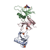
| Structure viewer | Molecule: MolmilJmol/JSmol |
|---|
- Downloads & links
-Download
| PDBx/mmCIF format | 3vnc.cif.gz | 71.3 KB | Display | PDBx/mmCIF format |
|---|---|---|---|---|
| PDB format | pdb3vnc.ent.gz | 53.6 KB | Display | PDB format |
| PDBx/mmJSON format | 3vnc.json.gz | Tree view | PDBx/mmJSON format | |
| Others |  Other downloads Other downloads |
-Validation report
| Arichive directory | https://data.pdbj.org/pub/pdb/validation_reports/vn/3vncftp://data.pdbj.org/pub/pdb/validation_reports/vn/3vnc | HTTPS FTP |
|---|
-Related structure data
| Similar structure data |
|---|


-Links
 PDBj
PDBj


- Assembly
| Deposited unit | 
| ||||||||
|---|---|---|---|---|---|---|---|---|---|
| 1 |
| ||||||||
| Unit cell |
|
-Components
| #1: Protein | Mass: 19223.031 Da / Num. of mol.: 2 / Fragment: Residues 25-192 Source method: isolated from a genetically manipulated source Source: (gene. exp.) Helicobacter pylori (bacteria) / Strain: 26695 / Gene: HP_0596 / Plasmid: pET22b / Production host: #2: Water | ChemComp-HOH / |  Mass: 18.015 Da / Num. of mol.: 55 / Source method: isolated from a natural source / Formula: H2O Mass: 18.015 Da / Num. of mol.: 55 / Source method: isolated from a natural source / Formula: H2O |
|---|
-Experimental details
-Experiment
| Experiment | Method: X-RAY DIFFRACTION / Number of used crystals: 1 |
|---|
- Sample preparation
| Crystal | Density Matthews: 3.32 Å3/Da / Density % sol: 62.9 % |
|---|---|
| Crystal grow | Temperature: 293 K / Method: vapor diffusion, hanging drop / pH: 4 Details: 16% PEG3350, 80% Tacsimate, 2% 1,2-propanediol, 5% MPD, 5% Glycerol, pH 4, VAPOR DIFFUSION, HANGING DROP, temperature 293K |
-Data collection
| Diffraction | Mean temperature: 90 K | ||||||||||||
|---|---|---|---|---|---|---|---|---|---|---|---|---|---|
| Diffraction source | Source: SYNCHROTRON / Site: Photon Factory  / Beamline: BL-17A / Wavelength: 0.97898, 0.97917, 0.96395 / Beamline: BL-17A / Wavelength: 0.97898, 0.97917, 0.96395 | ||||||||||||
| Detector | Type: ADSC QUANTUM 315r / Detector: CCD / Date: Oct 26, 2009 | ||||||||||||
| Radiation | Monochromator: Numerical link type Si(111) double crystal monochromator Protocol: MAD / Monochromatic (M) / Laue (L): M / Scattering type: x-ray | ||||||||||||
| Radiation wavelength |
| ||||||||||||
| Reflection | Resolution: 2.6→49.14 Å / Num. all: 15779 / Num. obs: 15054 / % possible obs: 95.4 % / Observed criterion σ(F): 0 / Observed criterion σ(I): 0 / Redundancy: 7.2 % / Biso Wilson estimate: 37.1 Å2 / Rmerge(I) obs: 0.102 / Rsym value: 0.102 / Net I/σ(I): 17.7 | ||||||||||||
| Reflection shell | Resolution: 2.6→2.74 Å / Redundancy: 7.3 % / Rmerge(I) obs: 0.374 / Mean I/σ(I) obs: 4.7 / Num. unique all: 2289 / Rsym value: 0.374 / % possible all: 99.9 |
- Processing
| Software |
| |||||||||||||||||||||||||
|---|---|---|---|---|---|---|---|---|---|---|---|---|---|---|---|---|---|---|---|---|---|---|---|---|---|---|
| Refinement | Method to determine structure: MAD / Resolution: 2.6→49.14 Å / Rfactor Rfree error: 0.007 / Data cutoff high absF: 2045408.21 / Data cutoff low absF: 0 / Isotropic thermal model: RESTRAINED / Cross valid method: THROUGHOUT / σ(F): 0 / σ(I): 0 / Stereochemistry target values: Engh & Huber / Details: BULK SOLVENT MODEL USED
| |||||||||||||||||||||||||
| Solvent computation | Solvent model: FLAT MODEL / Bsol: 58.6298 Å2 / ksol: 0.4 e/Å3 | |||||||||||||||||||||||||
| Displacement parameters | Biso mean: 48.7 Å2
| |||||||||||||||||||||||||
| Refine analyze |
| |||||||||||||||||||||||||
| Refinement step | Cycle: LAST / Resolution: 2.6→49.14 Å
| |||||||||||||||||||||||||
| Refine LS restraints |
| |||||||||||||||||||||||||
| LS refinement shell | Resolution: 2.6→2.76 Å / Rfactor Rfree error: 0.026 / Total num. of bins used: 6
| |||||||||||||||||||||||||
| Xplor file |
|
