- PDB-3vdx: Structure of a 16 nm protein cage designed by fusing symmetric ol... -
+
Open data
ID or keywords:
Loading...
-
Basic information
Entry
Database: PDB / ID: 3vdx
Title
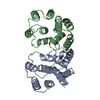
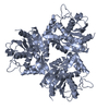
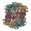
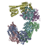
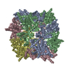
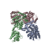
Structure of a 16 nm protein cage designed by fusing symmetric oligomeric domains
Components
Designed 16nm tetrahedral protein cage containing Non-haem bromoperoxidase BPO-A2 and Matrix protein 1
Keywords
DE NOVO PROTEIN / protein design / bionanotechnology / protein assembly / symmetry / biomaterials
Function / homology
Function and homology information
Assembly of Viral Components at the Budding Site / Influenza Infection / Fusion of the Influenza Virion to the Host Cell Endosome / Release / Budding / Packaging of Eight RNA Segments / Uncoating of the Influenza Virion / Entry of Influenza Virion into Host Cell via Endocytosis / Viral RNP Complexes in the Host Cell Nucleus / NEP/NS2 Interacts with the Cellular Export Machinery ...Assembly of Viral Components at the Budding Site / Influenza Infection / Fusion of the Influenza Virion to the Host Cell Endosome / Release / Budding / Packaging of Eight RNA Segments / Uncoating of the Influenza Virion / Entry of Influenza Virion into Host Cell via Endocytosis / Viral RNP Complexes in the Host Cell Nucleus / NEP/NS2 Interacts with the Cellular Export Machinery / Oxidoreductases; Acting on a peroxide as acceptor; Peroxidases / antibiotic biosynthetic process / Viral mRNA Translation / viral budding from plasma membrane / peroxidase activity / structural constituent of virion / host cell nucleus / virion membrane / RNA binding / extracellular region / plasma membrane Similarity search - Function
A: Designed 16nm tetrahedral protein cage containing Non-haem bromoperoxidase BPO-A2 and Matrix protein 1 B: Designed 16nm tetrahedral protein cage containing Non-haem bromoperoxidase BPO-A2 and Matrix protein 1 C: Designed 16nm tetrahedral protein cage containing Non-haem bromoperoxidase BPO-A2 and Matrix protein 1
A: Designed 16nm tetrahedral protein cage containing Non-haem bromoperoxidase BPO-A2 and Matrix protein 1 B: Designed 16nm tetrahedral protein cage containing Non-haem bromoperoxidase BPO-A2 and Matrix protein 1 C: Designed 16nm tetrahedral protein cage containing Non-haem bromoperoxidase BPO-A2 and Matrix protein 1
A: Designed 16nm tetrahedral protein cage containing Non-haem bromoperoxidase BPO-A2 and Matrix protein 1 B: Designed 16nm tetrahedral protein cage containing Non-haem bromoperoxidase BPO-A2 and Matrix protein 1 C: Designed 16nm tetrahedral protein cage containing Non-haem bromoperoxidase BPO-A2 and Matrix protein 1
A: Designed 16nm tetrahedral protein cage containing Non-haem bromoperoxidase BPO-A2 and Matrix protein 1 B: Designed 16nm tetrahedral protein cage containing Non-haem bromoperoxidase BPO-A2 and Matrix protein 1 C: Designed 16nm tetrahedral protein cage containing Non-haem bromoperoxidase BPO-A2 and Matrix protein 1
A: Designed 16nm tetrahedral protein cage containing Non-haem bromoperoxidase BPO-A2 and Matrix protein 1 B: Designed 16nm tetrahedral protein cage containing Non-haem bromoperoxidase BPO-A2 and Matrix protein 1 C: Designed 16nm tetrahedral protein cage containing Non-haem bromoperoxidase BPO-A2 and Matrix protein 1
In the structure databanks used in Yorodumi, some data are registered as the other names, "COVID-19 virus" and "2019-nCoV". Here are the details of the virus and the list of structure data.
Jan 31, 2019. EMDB accession codes are about to change! (news from PDBe EMDB page)
EMDB accession codes are about to change! (news from PDBe EMDB page)
The allocation of 4 digits for EMDB accession codes will soon come to an end. Whilst these codes will remain in use, new EMDB accession codes will include an additional digit and will expand incrementally as the available range of codes is exhausted. The current 4-digit format prefixed with “EMD-” (i.e. EMD-XXXX) will advance to a 5-digit format (i.e. EMD-XXXXX), and so on. It is currently estimated that the 4-digit codes will be depleted around Spring 2019, at which point the 5-digit format will come into force.
The EM Navigator/Yorodumi systems omit the EMD- prefix.
Related info.:Q: What is EMD? / ID/Accession-code notation in Yorodumi/EM Navigator
Yorodumi is a browser for structure data from EMDB, PDB, SASBDB, etc.
This page is also the successor to EM Navigator detail page, and also detail information page/front-end page for Omokage search.
The word "yorodu" (or yorozu) is an old Japanese word meaning "ten thousand". "mi" (miru) is to see.
Related info.:EMDB / PDB / SASBDB / Comparison of 3 databanks / Yorodumi Search / Aug 31, 2016. New EM Navigator & Yorodumi / Yorodumi Papers / Jmol/JSmol / Function and homology information / Changes in new EM Navigator and Yorodumi
 Movie
Movie Controller

 Yorodumi
 Open data
 Basic information
 Components
 Keywords
 Function and homology information
 Streptomyces aureofaciens (bacteria)
 Influenza A virus
 X-RAY DIFFRACTION /
  Authors
 Citation
 Structure visualization
 Downloads & links
 Other downloads
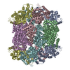


 PDBj
PDBj


 Assembly
 Sample preparation
 Processing