 Movie
Movie Controller

[English] 日本語
 Yorodumi
- PDB-3vb8: Crystal Structure of Engineered Protein, Northeast Structural Gen... -
+ Open data

- Basic information
| Entry | Database: PDB / ID: 3vb8 | ||||||
|---|---|---|---|---|---|---|---|
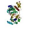
| Title | Crystal Structure of Engineered Protein, Northeast Structural Genomics Consortium Target OR43 | ||||||
 Components Components | Engineered protein | ||||||
 Keywords Keywords | DE NOVO PROTEIN / Structural Genomics / PSI-Biology / Northeast Structural Genomics Consortium / NESG | ||||||
| Function / homology | Immunoglobulin-like - #2930 / Immunoglobulin-like / Sandwich / Mainly Beta Function and homology information Function and homology information | ||||||
| Biological species | artificial gene (others) | ||||||
| Method |  X-RAY DIFFRACTION / SYNCHROTRON / MOLECULAR REPLACEMENT / Resolution: 2.9 Å X-RAY DIFFRACTION / SYNCHROTRON / MOLECULAR REPLACEMENT / Resolution: 2.9 Å | ||||||
 Authors Authors | Seetharaman, J. / Su, M. / Procko, E. / Baker, D. / Ciccosanti, C. / Sahdev, S. / Xiao, R. / Everett, J.K. / Acton, T.B. / Montelione, G.T. ...Seetharaman, J. / Su, M. / Procko, E. / Baker, D. / Ciccosanti, C. / Sahdev, S. / Xiao, R. / Everett, J.K. / Acton, T.B. / Montelione, G.T. / Hunt, J.F. / Tong, L. / Northeast Structural Genomics Consortium (NESG) | ||||||
 Citation Citation | Journal: J.Mol.Biol. / Year: 2013 Title: Computational design of a protein-based enzyme inhibitor. Authors: Procko, E. / Hedman, R. / Hamilton, K. / Seetharaman, J. / Fleishman, S.J. / Su, M. / Aramini, J. / Kornhaber, G. / Hunt, J.F. / Tong, L. / Montelione, G.T. / Baker, D. | ||||||
| History |
|
- Structure visualization
| Structure viewer | Molecule: MolmilJmol/JSmol |
|---|
- Downloads & links
-Download
| PDBx/mmCIF format | 3vb8.cif.gz | 77.7 KB | Display | PDBx/mmCIF format |
|---|---|---|---|---|
| PDB format | pdb3vb8.ent.gz | 58 KB | Display | PDB format |
| PDBx/mmJSON format | 3vb8.json.gz | Tree view | PDBx/mmJSON format | |
| Others |  Other downloads Other downloads |
-Validation report
| Arichive directory | https://data.pdbj.org/pub/pdb/validation_reports/vb/3vb8ftp://data.pdbj.org/pub/pdb/validation_reports/vb/3vb8 | HTTPS FTP |
|---|
-Related structure data
| Related structure data | 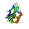 3ii3S S: Starting model for refinement |
|---|---|
| Similar structure data | |
| Other databases |


-Links
 PDBj
PDBj
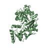
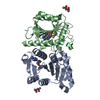
- Assembly
| Deposited unit | 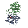
| ||||||||
|---|---|---|---|---|---|---|---|---|---|
| 1 |
| ||||||||
| 2 | 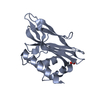
| ||||||||
| 3 | 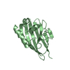
| ||||||||
| Unit cell |
|
-Components
| #1: Protein | Mass: 19221.904 Da / Num. of mol.: 2 Source method: isolated from a genetically manipulated source Source: (gene. exp.) artificial gene (others) / Production host:  #2: Chemical | ChemComp-SO4 / |   Mass: 96.063 Da / Num. of mol.: 1 / Source method: obtained synthetically / Formula: SO4 Mass: 96.063 Da / Num. of mol.: 1 / Source method: obtained synthetically / Formula: SO4#3: Water | ChemComp-HOH / |  Mass: 18.015 Da / Num. of mol.: 119 / Source method: isolated from a natural source / Formula: H2O Mass: 18.015 Da / Num. of mol.: 119 / Source method: isolated from a natural source / Formula: H2O |
|---|
-Experimental details
-Experiment
| Experiment | Method: X-RAY DIFFRACTION / Number of used crystals: 1 |
|---|
- Sample preparation
| Crystal | Density Matthews: 2.26 Å3/Da / Density % sol: 45.66 % |
|---|---|
| Crystal grow | Temperature: 293 K / pH: 8 Details: 1.59M MgSO4, 0.1 M Tris, pH 8, Microbatch under oil, temperature 293K |
-Data collection
| Diffraction | Mean temperature: 100 K |
|---|---|
| Diffraction source | Source: SYNCHROTRON / Site: NSLS  / Beamline: X4A / Wavelength: 0.979 Å / Beamline: X4A / Wavelength: 0.979 Å |
| Detector | Type: ADSC QUANTUM 4r / Detector: CCD / Date: Dec 10, 2001 |
| Radiation | Protocol: SINGLE WAVELENGTH / Monochromatic (M) / Laue (L): M / Scattering type: x-ray |
| Radiation wavelength | Wavelength: 0.979 Å / Relative weight: 1 |
| Reflection | Resolution: 2.8→50 Å / Num. all: 9162 / Num. obs: 9162 / % possible obs: 93.5 % / Observed criterion σ(F): 0 / Observed criterion σ(I): 0 / Redundancy: 7.3 % / Biso Wilson estimate: 13.8 Å2 / Rmerge(I) obs: 0.135 / Net I/σ(I): 10.4 |
| Reflection shell | Resolution: 2.8→2.9 Å / Num. unique all: 870 / % possible all: 98.3 |
- Processing
| Software |
| |||||||||||||||||||||||||
|---|---|---|---|---|---|---|---|---|---|---|---|---|---|---|---|---|---|---|---|---|---|---|---|---|---|---|
| Refinement | Method to determine structure: MOLECULAR REPLACEMENT Starting model: 3ii3 Resolution: 2.9→34.68 Å / Rfactor Rfree error: 0.009 / Data cutoff high absF: 92415.28 / Data cutoff low absF: 0 / Isotropic thermal model: RESTRAINED / Cross valid method: THROUGHOUT / σ(F): 2 / Stereochemistry target values: Engh & Huber / Details: BULK SOLVENT MODEL USED
| |||||||||||||||||||||||||
| Solvent computation | Solvent model: FLAT MODEL / Bsol: 37.1428 Å2 / ksol: 0.35 e/Å3 | |||||||||||||||||||||||||
| Displacement parameters | Biso mean: 48.9 Å2
| |||||||||||||||||||||||||
| Refine analyze |
| |||||||||||||||||||||||||
| Refinement step | Cycle: LAST / Resolution: 2.9→34.68 Å
| |||||||||||||||||||||||||
| Refine LS restraints |
| |||||||||||||||||||||||||
| Refine LS restraints NCS | NCS model details: NONE | |||||||||||||||||||||||||
| LS refinement shell | Resolution: 2.9→3.08 Å / Rfactor Rfree error: 0.029 / Total num. of bins used: 6
| |||||||||||||||||||||||||
| Xplor file |
|
