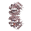
- PDB-3v48: Crystal Structure of the putative alpha/beta hydrolase RutD from ... -
+
Open data
ID or keywords:
Loading...
-
Basic information
Entry
Database: PDB / ID: 3v48
Title
Crystal Structure of the putative alpha/beta hydrolase RutD from E.coli
Components
Putative aminoacrylate hydrolase RutD
Keywords
HYDROLASE / Structural Genomics / PSI-Biology / New York Structural Genomics Research Consortium / NYSGRC
Function / homology
Function and homology information
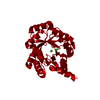
uracil catabolic process / nitrogen utilization / hydrolase activity, acting on carbon-nitrogen (but not peptide) bonds, in linear amides / Hydrolases; Acting on carbon-nitrogen bonds, other than peptide bonds; In linear amides Similarity search - Function
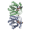
Mass: 29403.637 Da / Num. of mol.: 2 Source method: isolated from a genetically manipulated source Source: (gene. exp.) Escherichia coli SE11 (bacteria) / Strain: XL-1 Blue / Gene: ECSE_1071, rutD / Plasmid: pET15b, modified for TEV cleavage / Production host: Escherichia coli (E. coli) / Strain (production host): BL21DE3RIL References: UniProt: B6I985, Hydrolases; Acting on carbon-nitrogen bonds, other than peptide bonds; In linear amides
In the structure databanks used in Yorodumi, some data are registered as the other names, "COVID-19 virus" and "2019-nCoV". Here are the details of the virus and the list of structure data.
Jan 31, 2019. EMDB accession codes are about to change! (news from PDBe EMDB page)
EMDB accession codes are about to change! (news from PDBe EMDB page)
The allocation of 4 digits for EMDB accession codes will soon come to an end. Whilst these codes will remain in use, new EMDB accession codes will include an additional digit and will expand incrementally as the available range of codes is exhausted. The current 4-digit format prefixed with “EMD-” (i.e. EMD-XXXX) will advance to a 5-digit format (i.e. EMD-XXXXX), and so on. It is currently estimated that the 4-digit codes will be depleted around Spring 2019, at which point the 5-digit format will come into force.
The EM Navigator/Yorodumi systems omit the EMD- prefix.
Related info.:Q: What is EMD? / ID/Accession-code notation in Yorodumi/EM Navigator
Yorodumi is a browser for structure data from EMDB, PDB, SASBDB, etc.
This page is also the successor to EM Navigator detail page, and also detail information page/front-end page for Omokage search.
The word "yorodu" (or yorozu) is an old Japanese word meaning "ten thousand". "mi" (miru) is to see.
Related info.:EMDB / PDB / SASBDB / Comparison of 3 databanks / Yorodumi Search / Aug 31, 2016. New EM Navigator & Yorodumi / Yorodumi Papers / Jmol/JSmol / Function and homology information / Changes in new EM Navigator and Yorodumi
 Movie
Movie Controller

 Yorodumi
 Open data
 Basic information
 Components
 Keywords
 Function and homology information
 X-RAY DIFFRACTION /
  Authors
 Citation
 Structure visualization
 Downloads & links
 Other downloads
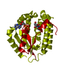


 PDBj
PDBj


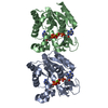
 Assembly
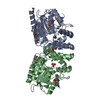

 Mass: 92.094 Da / Num. of mol.: 4 / Source method: obtained synthetically / Formula: C3H8O3
 Mass: 58.082 Da / Num. of mol.: 3 / Source method: obtained synthetically / Formula: CNS
 Mass: 18.015 Da / Num. of mol.: 313 / Source method: isolated from a natural source / Formula: H2O
 Sample preparation
 / Beamline: 21-ID-F / Wavelength: 0.9787 Å
 Processing