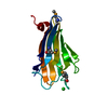
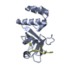
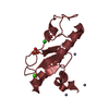
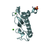
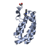
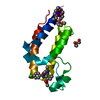
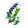
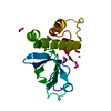
[English] 日本語
Yorodumi
- PDB-3v43: Crystal structure of MOZ -

+
Open data


ID or keywords:

Loading...

-
Basic information

Entry
Database: PDB / ID: 3v43
TitleCrystal structure of MOZ
Components
  • Histone H3.1Histone H3
  • Histone acetyltransferase KAT6A
KeywordsTRANSFERASE/STRUCTURAL PROTEIN / MOZ / PHD finger / Histone H3 / TRANSFERASE-STRUCTURAL PROTEIN complex
Function / homology
Function and homology information


histone H4K5 acetyltransferase activity / histone H4K8 acetyltransferase activity / histone H3K14 acetyltransferase activity / histone H4K12 acetyltransferase activity / histone H4K16 acetyltransferase activity / myeloid cell differentiation / regulation of developmental process / MOZ/MORF histone acetyltransferase complex / regulation of hemopoiesis / protein acetylation ...histone H4K5 acetyltransferase activity / histone H4K8 acetyltransferase activity / histone H3K14 acetyltransferase activity / histone H4K12 acetyltransferase activity / histone H4K16 acetyltransferase activity / myeloid cell differentiation / regulation of developmental process / MOZ/MORF histone acetyltransferase complex / regulation of hemopoiesis / protein acetylation / acetyltransferase activity / chromosome organization / Chromatin modifying enzymes / epigenetic regulation of gene expression / histone acetyltransferase activity / histone acetyltransferase / Regulation of TP53 Activity through Acetylation / telomere organization / RNA Polymerase I Promoter Opening / Interleukin-7 signaling / Assembly of the ORC complex at the origin of replication / DNA methylation / Condensation of Prophase Chromosomes / HCMV Late Events / Chromatin modifications during the maternal to zygotic transition (MZT) / ERCC6 (CSB) and EHMT2 (G9a) positively regulate rRNA expression / SIRT1 negatively regulates rRNA expression / PRC2 methylates histones and DNA / regulation of signal transduction by p53 class mediator / Defective pyroptosis / HDACs deacetylate histones / RNA Polymerase I Promoter Escape / transcription coregulator activity / Transcriptional regulation by small RNAs / Formation of the beta-catenin:TCF transactivating complex / RUNX1 regulates genes involved in megakaryocyte differentiation and platelet function / Activated PKN1 stimulates transcription of AR (androgen receptor) regulated genes KLK2 and KLK3 / NoRC negatively regulates rRNA expression / B-WICH complex positively regulates rRNA expression / HDMs demethylate histones / PML body / PKMTs methylate histone lysines / RMTs methylate histone arginines / Meiotic recombination / Pre-NOTCH Transcription and Translation / nucleosome assembly / Activation of anterior HOX genes in hindbrain development during early embryogenesis / HCMV Early Events / Transcriptional regulation of granulopoiesis / structural constituent of chromatin / nucleosome / cellular senescence / gene expression / RUNX1 regulates transcription of genes involved in differentiation of HSCs / chromatin organization / Factors involved in megakaryocyte development and platelet production / HATs acetylate histones / Senescence-Associated Secretory Phenotype (SASP) / Oxidative Stress Induced Senescence / DNA-binding transcription factor binding / Estrogen-dependent gene expression / transcription coactivator activity / nuclear speck / cadherin binding / Amyloid fiber formation / protein heterodimerization activity / negative regulation of DNA-templated transcription / positive regulation of gene expression / nucleolus / regulation of DNA-templated transcription / positive regulation of DNA-templated transcription / positive regulation of transcription by RNA polymerase II / protein-containing complex / DNA binding / extracellular exosome / zinc ion binding / extracellular region / nucleoplasm / membrane / nucleus / cytosol
Similarity search - Function
: / SAM domain-containing protein 1, WH domain / SAMD1-like winged helix (WH) domain profile. / MYST, zinc finger domain / MYST family zinc finger domain / Histone acetyltransferase domain, MYST-type / MOZ/SAS family / MYST-type histone acetyltransferase (HAT) domain profile. / Linker histone H1/H5, domain H15 / Linker histone H1/H5 globular (H15) domain profile. ...: / SAM domain-containing protein 1, WH domain / SAMD1-like winged helix (WH) domain profile. / MYST, zinc finger domain / MYST family zinc finger domain / Histone acetyltransferase domain, MYST-type / MOZ/SAS family / MYST-type histone acetyltransferase (HAT) domain profile. / Linker histone H1/H5, domain H15 / Linker histone H1/H5 globular (H15) domain profile. / Domain in histone families 1 and 5 / Zinc/RING finger domain, C3HC4 (zinc finger) / Herpes Virus-1 / PHD-finger / Acyl-CoA N-acyltransferase / Zinc finger PHD-type signature. / Zinc finger PHD-type profile. / Zinc finger, PHD-finger / Histone H3 signature 1. / Zinc finger, PHD-type / PHD zinc finger / Histone H3 signature 2. / Histone H3 / Histone H3/CENP-A / Histone H2A/H2B/H3 / Core histone H2A/H2B/H3/H4 / Zinc finger, FYVE/PHD-type / Histone-fold / Zinc finger, RING/FYVE/PHD-type / Winged helix DNA-binding domain superfamily / Winged helix-like DNA-binding domain superfamily / 2-Layer Sandwich / Alpha Beta
Similarity search - Domain/homology
ACETATE ION / Histone H3.1 / Histone acetyltransferase KAT6A
Similarity search - Component
Biological speciesHomo sapiens (human)
MethodX-RAY DIFFRACTION / SYNCHROTRON / SAD / Resolution: 1.47 Å
AuthorsQiu, Y. / Li, F.
CitationJournal: Genes Dev. / Year: 2012
Title: Combinatorial readout of unmodified H3R2 and acetylated H3K14 by the tandem PHD finger of MOZ reveals a regulatory mechanism for HOXA9 transcription
Authors: Qiu, Y. / Liu, L. / Zhao, C. / Han, C. / Li, F. / Zhang, J. / Wang, Y. / Li, G. / Mei, Y. / Wu, M. / Wu, J. / Shi, Y.
History
DepositionDec 14, 2011Deposition site: RCSB / Processing site: PDBJ
Revision 1.0Jun 27, 2012Provider: repository / Type: Initial release
Revision 1.1Jul 17, 2013Group: Database references
Revision 1.2Mar 20, 2024Group: Data collection / Database references / Derived calculations
Category: chem_comp_atom / chem_comp_bond ...chem_comp_atom / chem_comp_bond / database_2 / pdbx_struct_conn_angle / struct_conn / struct_ref_seq_dif / struct_site
Item: _database_2.pdbx_DOI / _database_2.pdbx_database_accession ..._database_2.pdbx_DOI / _database_2.pdbx_database_accession / _pdbx_struct_conn_angle.ptnr1_auth_comp_id / _pdbx_struct_conn_angle.ptnr1_auth_seq_id / _pdbx_struct_conn_angle.ptnr1_label_atom_id / _pdbx_struct_conn_angle.ptnr1_label_comp_id / _pdbx_struct_conn_angle.ptnr1_label_seq_id / _pdbx_struct_conn_angle.ptnr2_auth_seq_id / _pdbx_struct_conn_angle.ptnr2_label_asym_id / _pdbx_struct_conn_angle.ptnr3_auth_comp_id / _pdbx_struct_conn_angle.ptnr3_auth_seq_id / _pdbx_struct_conn_angle.ptnr3_label_atom_id / _pdbx_struct_conn_angle.ptnr3_label_comp_id / _pdbx_struct_conn_angle.ptnr3_label_seq_id / _pdbx_struct_conn_angle.value / _struct_conn.pdbx_dist_value / _struct_conn.ptnr1_auth_comp_id / _struct_conn.ptnr1_auth_seq_id / _struct_conn.ptnr1_label_asym_id / _struct_conn.ptnr1_label_atom_id / _struct_conn.ptnr1_label_comp_id / _struct_conn.ptnr1_label_seq_id / _struct_conn.ptnr2_auth_comp_id / _struct_conn.ptnr2_auth_seq_id / _struct_conn.ptnr2_label_asym_id / _struct_conn.ptnr2_label_atom_id / _struct_conn.ptnr2_label_comp_id / _struct_conn.ptnr2_label_seq_id / _struct_ref_seq_dif.details / _struct_site.pdbx_auth_asym_id / _struct_site.pdbx_auth_comp_id / _struct_site.pdbx_auth_seq_id

-
Structure visualization

Structure viewerMolecule:
MolmilJmol/JSmol

Downloads & links

-
Assembly

Deposited unit
A: Histone acetyltransferase KAT6A
Q: Histone H3.1
hetero molecules


Theoretical massNumber of molelcules
Total (without water)15,0917
Polymers14,7702
Non-polymers3215
Water2,828157
1


  • Idetical with deposited unit
  • defined by author&software
TypeNameSymmetry operationNumber
identity operation1_555x,y,z1
Buried area1240 Å2
ΔGint-2 kcal/mol
Surface area7390 Å2
MethodPISA
Unit cell
Length a, b, c (Å)35.080, 57.630, 76.510
Angle α, β, γ (deg.)90.000, 90.000, 90.000
Int Tables number19
Space group name H-MP212121

-
Components

#1: Protein Histone acetyltransferase KAT6A / MOZ / YBF2/SAS3 / SAS2 and TIP60 protein 3 / MYST-3 / Monocytic leukemia zinc finger protein / Runt- ...MOZ / YBF2/SAS3 / SAS2 and TIP60 protein 3 / MYST-3 / Monocytic leukemia zinc finger protein / Runt-related transcription factor-binding protein 2 / Zinc finger protein 220


Mass: 12779.969 Da / Num. of mol.: 1 / Fragment: UNP RESIDUES 204-313
Source method: isolated from a genetically manipulated source
Source: (gene. exp.) Homo sapiens (human) / Gene: KAT6A, MOZ, MYST3, RUNXBP2, ZNF220 / Plasmid: pET28a / Production host: Escherichia coli (E. coli) / Strain (production host): BL21(DE3) / References: UniProt: Q92794, histone acetyltransferase
#2: Protein/peptide Histone H3.1 / Histone H3 / Histone H3/a / Histone H3/b / Histone H3/c / Histone H3/d / Histone H3/f / Histone H3/h / Histone ...Histone H3/a / Histone H3/b / Histone H3/c / Histone H3/d / Histone H3/f / Histone H3/h / Histone H3/i / Histone H3/j / Histone H3/k / Histone H3/l


Mass: 1990.315 Da / Num. of mol.: 1 / Fragment: UNP RESIDUES 2-19 / Source method: obtained synthetically / Details: The peptide was chemically synthesized. / Source: (synth.) Homo sapiens (human) / References: UniProt: P68431
#3: Chemical
ChemComp-ZN / ZINC ION


Mass: 65.409 Da / Num. of mol.: 4 / Source method: obtained synthetically / Formula: Zn
#4: Chemical ChemComp-ACT / ACETATE ION / Acetate


Mass: 59.044 Da / Num. of mol.: 1 / Source method: obtained synthetically / Formula: C2H3O2
#5: Water ChemComp-HOH / water / Water


Mass: 18.015 Da / Num. of mol.: 157 / Source method: isolated from a natural source / Formula: H2O

-
Experimental details

-
Experiment

ExperimentMethod: X-RAY DIFFRACTION / Number of used crystals: 1

-
Sample preparation

CrystalDensity Matthews: 2.62 Å3/Da / Density % sol: 53.02 % / Mosaicity: 0.46 °
Crystal growTemperature: 293 K / Method: vapor diffusion, hanging drop / pH: 5.6
Details: 0.2M ammonium acetate, 0.1M tri-sodium citrate, 30% w/v PEG 4000, pH 5.6, vapor diffusion, hanging drop, temperature 293K

-
Data collection

DiffractionMean temperature: 100 K
Diffraction sourceSource: SYNCHROTRON / Site: SSRF / Beamline: BL17U / Wavelength: 0.9793 Å
DetectorType: RAYONIX MX-225 / Detector: CCD / Date: May 30, 2011
RadiationProtocol: SINGLE WAVELENGTH / Monochromatic (M) / Laue (L): M / Scattering type: x-ray
Radiation wavelengthWavelength: 0.9793 Å / Relative weight: 1
ReflectionRedundancy: 13.6 % / Av σ(I) over netI: 7.6 / Rsym value: 0.063 / D res high: 1.467 Å / D res low: 38.255 Å / Num. obs: 27192 / % possible obs: 99.6
Diffraction reflection shell
Highest resolution (Å)Lowest resolution (Å)% possible obs (%)IDRmerge(I) obsRsym valueRedundancy
1.471.5597.810.4560.45612.9
1.551.6410010.2820.28214
1.641.7510010.1760.17614
1.751.8910010.1160.11614
1.892.0710010.0890.08913.9
2.072.3210010.0590.05913.8
2.322.6899.910.0510.05113.6
2.683.2899.610.0580.05813.3
3.284.6499.610.0390.03912.4
4.6438.2698.110.0350.03512.3
ReflectionResolution: 1.467→38.255 Å / Num. all: 27192 / Num. obs: 27192 / % possible obs: 99.6 % / Observed criterion σ(F): -3 / Observed criterion σ(I): 0 / Redundancy: 13.6 % / Rsym value: 0.063 / Net I/σ(I): 24
Reflection shell

Diffraction-ID: 1

Resolution (Å)Redundancy (%)Rmerge(I) allRmerge(I) obsMean I/σ(I) obsNum. measured allNum. unique allRpim(I) allRrim(I) allRsym valueNet I/σ(I) obs% possible all
1.47-1.5512.90.4930.4561.64920738200.1350.4930.4565.897.8
1.55-1.64140.3060.2822.65199837020.0810.3060.2829.5100
1.64-1.75140.1930.1764.14925835220.0510.1930.17614.1100
1.75-1.89140.1320.11664539732430.0350.1320.11619.6100
1.89-2.0713.90.1040.0896.94224630420.0280.1040.08926.3100
2.07-2.3213.80.0720.059113806727560.0190.0720.05933.6100
2.32-2.6813.60.0660.051123312124280.0180.0660.05137.199.9
2.68-3.2813.30.070.0589.82746020720.0190.070.05843.399.6
3.28-4.6412.40.0490.03915.82036116440.0140.0490.0395099.6
4.64-38.25512.30.0510.03517.8118039630.0140.0510.03547.898.1

-
Phasing

PhasingMethod: SAD

-
Processing

Software
NameVersionClassificationNB
MOSFLMdata reduction
SCALA3.3.9data scaling
SHELXphasing
REFMAC5.5.0109refinement
PDB_EXTRACT3.1data extraction
MAR345dtbdata collection
SHELXDphasing
RefinementMethod to determine structure: SAD / Resolution: 1.47→19.13 Å / Cor.coef. Fo:Fc: 0.966 / Cor.coef. Fo:Fc free: 0.957 / Occupancy max: 1 / Occupancy min: 0 / SU B: 1.658 / SU ML: 0.03 / Cross valid method: THROUGHOUT / σ(F): 0 / ESU R Free: 0.055 / Stereochemistry target values: MAXIMUM LIKELIHOOD
Details: HYDROGENS HAVE BEEN ADDED IN THE RIDING POSITIONS U VALUES: REFINED INDIVIDUALLY
RfactorNum. reflection% reflectionSelection details
Rfree0.1762 1354 5.1 %RANDOM
Rwork0.1529 ---
obs0.1541 26750 98.44 %-
all-27138 --
Solvent computationIon probe radii: 0.8 Å / Shrinkage radii: 0.8 Å / VDW probe radii: 1.4 Å / Solvent model: MASK
Displacement parametersBiso max: 127.42 Å2 / Biso mean: 20.4177 Å2 / Biso min: 8.85 Å2
Baniso -1Baniso -2Baniso -3
1--0.02 Å20 Å20 Å2
2---0.04 Å20 Å2
3---0.06 Å2
Refinement stepCycle: LAST / Resolution: 1.47→19.13 Å
ProteinNucleic acidLigandSolventTotal
Num. atoms913 0 8 157 1078
Refine LS restraints
Refine-IDTypeDev idealDev ideal targetNumber
X-RAY DIFFRACTIONr_bond_refined_d0.010.022942
X-RAY DIFFRACTIONr_angle_refined_deg1.3861.9741272
X-RAY DIFFRACTIONr_dihedral_angle_1_deg5.3925123
X-RAY DIFFRACTIONr_dihedral_angle_2_deg31.90222.82139
X-RAY DIFFRACTIONr_dihedral_angle_3_deg13.24215168
X-RAY DIFFRACTIONr_dihedral_angle_4_deg21.5691510
X-RAY DIFFRACTIONr_chiral_restr0.0910.2137
X-RAY DIFFRACTIONr_gen_planes_refined0.0060.022718
X-RAY DIFFRACTIONr_mcbond_it1.2121.5609
X-RAY DIFFRACTIONr_mcangle_it2.0382982
X-RAY DIFFRACTIONr_scbond_it2.8923333
X-RAY DIFFRACTIONr_scangle_it4.3964.5287
X-RAY DIFFRACTIONr_rigid_bond_restr1.2133942
LS refinement shellResolution: 1.467→1.505 Å / Total num. of bins used: 20
RfactorNum. reflection% reflection
Rfree0.242 87 -
Rwork0.206 1683 -
all-1770 -
obs--93.9 %

+
About Yorodumi

-
News

-
Feb 9, 2022. New format data for meta-information of EMDB entries

New format data for meta-information of EMDB entries

  • Version 3 of the EMDB header file is now the official format.
  • The previous official version 1.9 will be removed from the archive.

Related info.:EMDB header

External links:wwPDB to switch to version 3 of the EMDB data model

-
Aug 12, 2020. Covid-19 info

Covid-19 info

URL: https://pdbj.org/emnavi/covid19.php

New page: Covid-19 featured information page in EM Navigator.

Related info.:Covid-19 info / Mar 5, 2020. Novel coronavirus structure data

+
Mar 5, 2020. Novel coronavirus structure data

Novel coronavirus structure data

Related info.:Yorodumi Speices / Aug 12, 2020. Covid-19 info

External links:COVID-19 featured content - PDBj / Molecule of the Month (242):Coronavirus Proteases

+
Jan 31, 2019. EMDB accession codes are about to change! (news from PDBe EMDB page)

EMDB accession codes are about to change! (news from PDBe EMDB page)

  • The allocation of 4 digits for EMDB accession codes will soon come to an end. Whilst these codes will remain in use, new EMDB accession codes will include an additional digit and will expand incrementally as the available range of codes is exhausted. The current 4-digit format prefixed with “EMD-” (i.e. EMD-XXXX) will advance to a 5-digit format (i.e. EMD-XXXXX), and so on. It is currently estimated that the 4-digit codes will be depleted around Spring 2019, at which point the 5-digit format will come into force.
  • The EM Navigator/Yorodumi systems omit the EMD- prefix.

Related info.:Q: What is EMD? / ID/Accession-code notation in Yorodumi/EM Navigator

External links:EMDB Accession Codes are Changing Soon! / Contact to PDBj

+
Jul 12, 2017. Major update of PDB

Major update of PDB

  • wwPDB released updated PDB data conforming to the new PDBx/mmCIF dictionary.
  • This is a major update changing the version number from 4 to 5, and with Remediation, in which all the entries are updated.
  • In this update, many items about electron microscopy experimental information are reorganized (e.g. em_software).
  • Now, EM Navigator and Yorodumi are based on the updated data.

External links:wwPDB Remediation / Enriched Model Files Conforming to OneDep Data Standards Now Available in the PDB FTP Archive

-
Yorodumi

Thousand views of thousand structures

  • Yorodumi is a browser for structure data from EMDB, PDB, SASBDB, etc.
  • This page is also the successor to EM Navigator detail page, and also detail information page/front-end page for Omokage search.
  • The word "yorodu" (or yorozu) is an old Japanese word meaning "ten thousand". "mi" (miru) is to see.

Related info.:EMDB / PDB / SASBDB / Comparison of 3 databanks / Yorodumi Search / Aug 31, 2016. New EM Navigator & Yorodumi / Yorodumi Papers / Jmol/JSmol / Function and homology information / Changes in new EM Navigator and Yorodumi

Read more