 Movie
Movie Controller

[English] 日本語
 Yorodumi
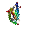
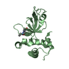
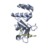
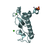
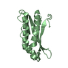
- PDB-1khc: Crystal Structure of the PWWP Domain of Mammalian DNA Methyltrans... -
+ Open data

- Basic information
| Entry | Database: PDB / ID: 1khc | ||||||
|---|---|---|---|---|---|---|---|
| Title | Crystal Structure of the PWWP Domain of Mammalian DNA Methyltransferase Dnmt3b | ||||||
 Components Components | DNA cytosine-5 methyltransferase 3B2 | ||||||
 Keywords Keywords | TRANSFERASE / Five beta-sheets barrel followed by Five-helix bundle | ||||||
| Function / homology |  Function and homology information Function and homology informationgene silencing by piRNA-directed DNA methylation / DNA (cytosine-5-)-methyltransferase activity, acting on CpN substrates / autosome genomic imprinting / SUMOylation of DNA methylation proteins / epigenetic programming of gene expression / PRC2 methylates histones and DNA / DNA (cytosine-5-)-methyltransferase activity, acting on CpG substrates / transposable element silencing by heterochromatin formation / DNA (cytosine-5-)-methyltransferase / DNA (cytosine-5-)-methyltransferase activity ...gene silencing by piRNA-directed DNA methylation / DNA (cytosine-5-)-methyltransferase activity, acting on CpN substrates / autosome genomic imprinting / SUMOylation of DNA methylation proteins / epigenetic programming of gene expression / PRC2 methylates histones and DNA / DNA (cytosine-5-)-methyltransferase activity, acting on CpG substrates / transposable element silencing by heterochromatin formation / DNA (cytosine-5-)-methyltransferase / DNA (cytosine-5-)-methyltransferase activity / protein-containing complex localization / DNA methylation-dependent constitutive heterochromatin formation / lncRNA binding / negative regulation of gene expression, epigenetic / chromosome, centromeric region / catalytic complex / heterochromatin / positive regulation of neuron differentiation / epigenetic regulation of gene expression / methyltransferase activity / cellular response to amino acid stimulus / histone deacetylase binding / transcription corepressor activity / heterochromatin formation / methylation / negative regulation of DNA-templated transcription / chromatin binding / positive regulation of gene expression / negative regulation of transcription by RNA polymerase II / DNA binding / zinc ion binding / nucleoplasm / nucleus Similarity search - Function | ||||||
| Biological species |  | ||||||
| Method |  X-RAY DIFFRACTION / MAD / Resolution: 1.8 Å X-RAY DIFFRACTION / MAD / Resolution: 1.8 Å | ||||||
 Authors Authors | Qiu, C. / Sawada, K. / Zhang, X. / Cheng, X. | ||||||
 Citation Citation | Journal: Nat.Struct.Biol. / Year: 2002 Title: The PWWP domain of mammalian DNA methyltransferase Dnmt3b defines a new family of DNA-binding folds. Authors: Qiu, C. / Sawada, K. / Zhang, X. / Cheng, X. | ||||||
| History |
| ||||||
| Remark 600 | HETEROGEN THE AUTHORS WERE UNABLE TO FIGURE OUT THE IDENTITY OF THE MOLECULES LABELED AS "UNX" FROM ...HETEROGEN THE AUTHORS WERE UNABLE TO FIGURE OUT THE IDENTITY OF THE MOLECULES LABELED AS "UNX" FROM THE ELECTRON DENSITY. |
- Structure visualization
| Structure viewer | Molecule: MolmilJmol/JSmol |
|---|
- Downloads & links
-Download
| PDBx/mmCIF format | 1khc.cif.gz | 44.4 KB | Display | PDBx/mmCIF format |
|---|---|---|---|---|
| PDB format | pdb1khc.ent.gz | 30.4 KB | Display | PDB format |
| PDBx/mmJSON format | 1khc.json.gz | Tree view | PDBx/mmJSON format | |
| Others |  Other downloads Other downloads |
-Validation report
| Arichive directory | https://data.pdbj.org/pub/pdb/validation_reports/kh/1khcftp://data.pdbj.org/pub/pdb/validation_reports/kh/1khc | HTTPS FTP |
|---|
-Related structure data
| Similar structure data |
|---|


-Links
 PDBj
PDBj
- Assembly
| Deposited unit | 
| ||||||||||||
|---|---|---|---|---|---|---|---|---|---|---|---|---|---|
| 1 |
| ||||||||||||
| Unit cell |
| ||||||||||||
| Components on special symmetry positions |
|
-Components
| #1: Protein | Mass: 16670.039 Da / Num. of mol.: 1 / Fragment: PWWP domain (residues 219-365) Source method: isolated from a genetically manipulated source Source: (gene. exp.)  References: UniProt: O88509, DNA (cytosine-5-)-methyltransferase | ||
|---|---|---|---|
| #2: Chemical | ChemComp-UNX /   Num. of mol.: 22 / Source method: obtained synthetically Num. of mol.: 22 / Source method: obtained synthetically#3: Water | ChemComp-HOH / |  Mass: 18.015 Da / Num. of mol.: 162 / Source method: isolated from a natural source / Formula: H2O Mass: 18.015 Da / Num. of mol.: 162 / Source method: isolated from a natural source / Formula: H2O |
-Experimental details
-Experiment
| Experiment | Method: X-RAY DIFFRACTION / Number of used crystals: 1 |
|---|
- Sample preparation
| Crystal | Density Matthews: 2.25 Å3/Da / Density % sol: 48 % | |||||||||||||||||||||||||||||||||||||||||||||||||||||||||||||||
|---|---|---|---|---|---|---|---|---|---|---|---|---|---|---|---|---|---|---|---|---|---|---|---|---|---|---|---|---|---|---|---|---|---|---|---|---|---|---|---|---|---|---|---|---|---|---|---|---|---|---|---|---|---|---|---|---|---|---|---|---|---|---|---|---|
| Crystal grow | Temperature: 289 K / Method: vapor diffusion, hanging drop / pH: 6.5 Details: 30% PEG 8000, 0.1M sodium cacodylate, 0.2M sodium acetate, pH 6.5, VAPOR DIFFUSION, HANGING DROP, temperature 289K | |||||||||||||||||||||||||||||||||||||||||||||||||||||||||||||||
| Crystal grow | *PLUS Temperature: 16 ℃ / pH: 7 | |||||||||||||||||||||||||||||||||||||||||||||||||||||||||||||||
| Components of the solutions | *PLUS
|
-Data collection
| Diffraction |
| |||||||||
|---|---|---|---|---|---|---|---|---|---|---|
| Diffraction source | Source: ROTATING ANODE / Type: RIGAKU / Wavelength: 1.54 Å | |||||||||
| Detector | Type: RIGAKU RAXIS IV / Detector: IMAGE PLATE / Date: May 27, 2000 | |||||||||
| Radiation | Monochromator: confocal multilayer mirror / Protocol: SINGLE WAVELENGTH / Monochromatic (M) / Laue (L): M / Scattering type: x-ray | |||||||||
| Radiation wavelength | Wavelength: 1.54 Å / Relative weight: 1 | |||||||||
| Reflection | Resolution: 1.8→20 Å / Num. all: 15308 / Num. obs: 15308 / % possible obs: 99.2 % / Observed criterion σ(F): 0 / Observed criterion σ(I): 0 / Redundancy: 9.5 % / Rmerge(I) obs: 0.038 / Net I/σ(I): 23.6 | |||||||||
| Reflection shell | Resolution: 1.8→1.83 Å / Redundancy: 4.3 % / Rmerge(I) obs: 0.148 / Mean I/σ(I) obs: 9.4 / % possible all: 94.3 | |||||||||
| Reflection | *PLUS Num. measured all: 145433 | |||||||||
| Reflection shell | *PLUS % possible obs: 94.3 % |
- Processing
| Software |
| ||||||||||||||||||||
|---|---|---|---|---|---|---|---|---|---|---|---|---|---|---|---|---|---|---|---|---|---|
| Refinement | Method to determine structure: MAD / Resolution: 1.8→20 Å / Data cutoff high absF: 1000000 / Data cutoff low absF: 0.001 / σ(F): 2
| ||||||||||||||||||||
| Displacement parameters | Biso mean: 15.442 Å2 | ||||||||||||||||||||
| Refinement step | Cycle: LAST / Resolution: 1.8→20 Å
| ||||||||||||||||||||
| Refine LS restraints |
| ||||||||||||||||||||
| LS refinement shell | Resolution: 1.8→1.88 Å / Total num. of bins used: 8
| ||||||||||||||||||||
| Refinement | *PLUS Lowest resolution: 20 Å / % reflection Rfree: 10 % / Rfactor obs: 0.19 / Rfactor Rwork: 0.19 | ||||||||||||||||||||
| Solvent computation | *PLUS | ||||||||||||||||||||
| Displacement parameters | *PLUS | ||||||||||||||||||||
| Refine LS restraints | *PLUS
| ||||||||||||||||||||
| LS refinement shell | *PLUS Highest resolution: 1.8 Å / Rfactor Rfree: 0.282 / % reflection Rfree: 11.4 % / Rfactor Rwork: 0.275 |
