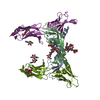
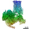
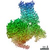
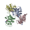
[English] 日本語
Yorodumi
- PDB-3v2a: VEGFR-2/VEGF-A COMPLEX STRUCTURE -

+
Open data


ID or keywords:

Loading...

-
Basic information

Entry
Database: PDB / ID: 3v2a
TitleVEGFR-2/VEGF-A COMPLEX STRUCTURE
Components
  • Vascular endothelial growth factor A
  • Vascular endothelial growth factor receptor 2VEGF receptor
KeywordsHORMONE/SIGNALING PROTEIN / IG-HOMOLOGY DOMAIN / VEGFR-2 / GROWTH FACTOR RECEPTOR / VEGF LIGAND / HORMONE-SIGNALING PROTEIN COMPLEX / ANGIOGENESIS / MEMBRANE / VEGF-A
Function / homology
Function and homology information


basophil chemotaxis / cellular stress response to acid chemical / VEGF-A complex / Signaling by VEGF / lymph vessel morphogenesis / vascular endothelial growth factor receptor 1 binding / negative regulation of establishment of endothelial barrier / vascular endothelial growth factor receptor binding / blood vessel endothelial cell differentiation / cellular response to hydrogen sulfide ...basophil chemotaxis / cellular stress response to acid chemical / VEGF-A complex / Signaling by VEGF / lymph vessel morphogenesis / vascular endothelial growth factor receptor 1 binding / negative regulation of establishment of endothelial barrier / vascular endothelial growth factor receptor binding / blood vessel endothelial cell differentiation / cellular response to hydrogen sulfide / VEGF ligand-receptor interactions / positive regulation of mast cell chemotaxis / positive regulation of peptidyl-tyrosine autophosphorylation / negative regulation of adherens junction organization / post-embryonic camera-type eye development / primitive erythrocyte differentiation / regulation of bone development / positive regulation of protein kinase C signaling / negative regulation of blood-brain barrier permeability / positive regulation of cell proliferation by VEGF-activated platelet derived growth factor receptor signaling pathway / VEGF-activated neuropilin signaling pathway / positive regulation of vascular endothelial growth factor signaling pathway / regulation of hematopoietic progenitor cell differentiation / Signaling by membrane-tethered fusions of PDGFRA or PDGFRB / coronary vein morphogenesis / cardiac vascular smooth muscle cell development / endothelium development / motor neuron migration / Neurophilin interactions with VEGF and VEGFR / positive regulation of trophoblast cell migration / endothelial cell chemotaxis / vascular endothelial growth factor binding / positive regulation of endothelial cell chemotaxis by VEGF-activated vascular endothelial growth factor receptor signaling pathway / eye photoreceptor cell development / vascular endothelial growth factor receptor-2 signaling pathway / VEGF binds to VEGFR leading to receptor dimerization / regulation of nitric oxide mediated signal transduction / endocardium development / positive regulation of axon extension involved in axon guidance / positive regulation of cell migration by vascular endothelial growth factor signaling pathway / positive regulation of branching involved in ureteric bud morphogenesis / positive regulation of protein localization to early endosome / vascular wound healing / vascular endothelial growth factor receptor activity / positive regulation of blood vessel endothelial cell proliferation involved in sprouting angiogenesis / post-embryonic camera-type eye morphogenesis / endothelial cell differentiation / mesenchymal cell proliferation / positive regulation of vasculogenesis / lymph vessel development / tube formation / camera-type eye morphogenesis / positive regulation of epithelial tube formation / negative regulation of cell-cell adhesion mediated by cadherin / neuropilin binding / induction of positive chemotaxis / coronary artery morphogenesis / vascular endothelial growth factor receptor 2 binding / commissural neuron axon guidance / positive regulation of vascular permeability / dopaminergic neuron differentiation / positive regulation of BMP signaling pathway / surfactant homeostasis / positive regulation of endothelial cell chemotaxis / platelet-derived growth factor receptor binding / epithelial cell maturation / extracellular matrix binding / anchoring junction / cell migration involved in sprouting angiogenesis / retinal ganglion cell axon guidance / artery morphogenesis / cardiac muscle cell development / sprouting angiogenesis / positive regulation of positive chemotaxis / embryonic hemopoiesis / Regulation of gene expression by Hypoxia-inducible Factor / vascular endothelial growth factor signaling pathway / positive regulation of leukocyte migration / positive regulation of p38MAPK cascade / positive regulation of mesenchymal cell proliferation / positive regulation of mitochondrial depolarization / positive regulation of DNA biosynthetic process / positive regulation of mitochondrial fission / branching involved in blood vessel morphogenesis / positive regulation of neuroblast proliferation / positive regulation of cell migration involved in sprouting angiogenesis / lung alveolus development / positive chemotaxis / transmembrane receptor protein tyrosine kinase activator activity / positive regulation of stem cell proliferation / positive regulation of sprouting angiogenesis / outflow tract morphogenesis / activation of protein kinase activity / chemoattractant activity / positive regulation of nitric-oxide synthase biosynthetic process / growth factor binding / sorting endosome / positive regulation of focal adhesion assembly / regulation of MAPK cascade / mesoderm development
Similarity search - Function
Vascular endothelial growth factor, heparin-binding domain / Vascular endothelial growth factor, heparin-binding domain superfamily / VEGF heparin-binding domain / Vascular endothelial growth factor receptor 2 (VEGFR2) / VEGFR-2, transmembrane domain / VEGFR-2 Transmembrane domain / Vascular endothelial growth factor receptor 1-like, Ig-like domain / PDGF/VEGF domain / Platelet-derived growth factor, conserved site / PDGF/VEGF domain ...Vascular endothelial growth factor, heparin-binding domain / Vascular endothelial growth factor, heparin-binding domain superfamily / VEGF heparin-binding domain / Vascular endothelial growth factor receptor 2 (VEGFR2) / VEGFR-2, transmembrane domain / VEGFR-2 Transmembrane domain / Vascular endothelial growth factor receptor 1-like, Ig-like domain / PDGF/VEGF domain / Platelet-derived growth factor, conserved site / PDGF/VEGF domain / Platelet-derived growth factor (PDGF) family signature. / Platelet-derived growth factor (PDGF) family profile. / Platelet-derived and vascular endothelial growth factors (PDGF, VEGF) family / Tyrosine-protein kinase, receptor class III, conserved site / Receptor tyrosine kinase class III signature. / Cystine Knot Cytokines, subunit B / Cystine-knot cytokines / Immunoglobulin domain / Cystine-knot cytokine / Immunoglobulin / Immunoglobulin domain / Immunoglobulin I-set / Immunoglobulin I-set domain / Immunoglobulin subtype 2 / Immunoglobulin C-2 Type / Ribbon / Tyrosine-protein kinase, catalytic domain / Tyrosine kinase, catalytic domain / Tyrosine protein kinases specific active-site signature. / Immunoglobulin subtype / Immunoglobulin / Tyrosine-protein kinase, active site / Protein tyrosine and serine/threonine kinase / Serine-threonine/tyrosine-protein kinase, catalytic domain / Ig-like domain profile. / Immunoglobulin-like domain / Immunoglobulin-like domain superfamily / Immunoglobulins / Protein kinase, ATP binding site / Protein kinases ATP-binding region signature. / Immunoglobulin-like fold / Protein kinase domain profile. / Protein kinase domain / Protein kinase-like domain superfamily / Immunoglobulin-like / Sandwich / Mainly Beta
Similarity search - Domain/homology
Vascular endothelial growth factor A, long form / Vascular endothelial growth factor receptor 2
Similarity search - Component
Biological speciesHomo sapiens (human)
MethodX-RAY DIFFRACTION / SYNCHROTRON / MOLECULAR REPLACEMENT / Resolution: 3.204 Å
AuthorsBrozzo, M.S. / Leppanen, V.-M. / Winkler, F.K. / Ballmer-Hofer, K.
CitationJournal: Blood / Year: 2012
Title: Thermodynamic and structural description of allosterically regulated VEGFR-2 dimerization.
Authors: Brozzo, M.S. / Bjelic, S. / Kisko, K. / Schleier, T. / Leppanen, V.M. / Alitalo, K. / Winkler, F.K. / Ballmer-Hofer, K.
History
DepositionDec 12, 2011Deposition site: RCSB / Processing site: RCSB
Revision 1.0Jan 18, 2012Provider: repository / Type: Initial release
Revision 1.1Mar 7, 2012Group: Database references
Revision 1.2Nov 8, 2017Group: Refinement description / Category: software / Item: _software.name
Revision 1.3Apr 3, 2024Group: Data collection / Database references / Refinement description
Category: chem_comp_atom / chem_comp_bond ...chem_comp_atom / chem_comp_bond / database_2 / pdbx_initial_refinement_model / struct_ref_seq_dif
Item: _database_2.pdbx_DOI / _database_2.pdbx_database_accession / _struct_ref_seq_dif.details

-
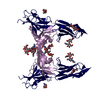
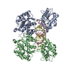
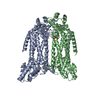
Structure visualization

Structure viewerMolecule:
MolmilJmol/JSmol

Downloads & links

-
Assembly

Deposited unit
R: Vascular endothelial growth factor receptor 2
A: Vascular endothelial growth factor A


Theoretical massNumber of molelcules
Total (without water)102,1092
Polymers102,1092
Non-polymers00
Water0
1
R: Vascular endothelial growth factor receptor 2
A: Vascular endothelial growth factor A

R: Vascular endothelial growth factor receptor 2
A: Vascular endothelial growth factor A


Theoretical massNumber of molelcules
Total (without water)204,2184
Polymers204,2184
Non-polymers00
Water0
TypeNameSymmetry operationNumber
identity operation1_555x,y,z1
crystal symmetry operation10_555-y,-x,-z+1/61
Buried area6960 Å2
ΔGint-57 kcal/mol
Surface area27100 Å2
MethodPISA
Unit cell
Length a, b, c (Å)80.969, 80.969, 332.063
Angle α, β, γ (deg.)90.00, 90.00, 120.00
Int Tables number179
Space group name H-MP6522
Detailsthe asymmetric unit contains one copy of each component of the complex

-
Components

#1: Protein Vascular endothelial growth factor receptor 2 / VEGF receptor / VEGFR-2 / Fetal liver kinase 1 / FLK-1 / Kinase insert domain receptor / KDR / Protein-tyrosine ...VEGFR-2 / Fetal liver kinase 1 / FLK-1 / Kinase insert domain receptor / KDR / Protein-tyrosine kinase receptor flk-1


Mass: 86566.461 Da / Num. of mol.: 1 / Fragment: UNP residues 1-764
Source method: isolated from a genetically manipulated source
Source: (gene. exp.) Homo sapiens (human) / Gene: FLK1, KDR, VEGFR2 / Production host: 
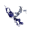
Spodoptera frugiperda (fall armyworm) / Strain (production host): Sf21
References: UniProt: P35968, receptor protein-tyrosine kinase
#2: Protein Vascular endothelial growth factor A / / VEGF-A / Vascular permeability factor / VPF


Mass: 15542.596 Da / Num. of mol.: 1 / Fragment: UNP residues 27-140
Source method: isolated from a genetically manipulated source
Source: (gene. exp.) Homo sapiens (human) / Gene: VEGF, VEGF-A, VEGFA / Production host: Pichia pastoris (fungus) / References: UniProt: P15692

-
Experimental details

-
Experiment

ExperimentMethod: X-RAY DIFFRACTION / Number of used crystals: 1

-
Sample preparation

Crystal growTemperature: 293 K / Method: vapor diffusion, sitting drop / pH: 7.5
Details: 0.1 M NaCl, 1.6 M ammonium sulfate, pH 7.5, VAPOR DIFFUSION, SITTING DROP, temperature 293K

-
Data collection

DiffractionMean temperature: 100 K
Diffraction sourceSource: SYNCHROTRON / Site: SLS / Beamline: X06SA / Wavelength: 0.9999 Å
DetectorType: DECTRIS PILATUS 6M / Detector: PIXEL / Details: DYNAMICALLY BENDABLE MIRROR
RadiationMonochromator: LN2 COOLED FIXED EXIT SI(111) / Protocol: SINGLE WAVELENGTH / Monochromatic (M) / Laue (L): M / Scattering type: x-ray
Radiation wavelengthWavelength: 0.9999 Å / Relative weight: 1
ReflectionResolution: 3.2→68.608 Å / % possible obs: 95.9 % / Observed criterion σ(I): -3 / Redundancy: 2.1 %
Reflection shellResolution: 3.2→3.4 Å / Redundancy: 2.1 % / Mean I/σ(I) obs: 1.25 / Rsym value: 0.76 / % possible all: 94.1

-
Processing

Software
NameVersionClassification
RemDAqdata collection
PHASERphasing
PHENIX(phenix.refine: 1.7_650)refinement
XDSdata reduction
XSCALEdata scaling
RefinementMethod to determine structure: MOLECULAR REPLACEMENT
Starting model: VEGF-E and VEGFR-2 VEGF-C complex structure

Resolution: 3.204→68.608 Å / SU ML: 0.35 / Isotropic thermal model: isotropic / Cross valid method: THROUGHOUT / σ(F): 2 / Phase error: 30 / Stereochemistry target values: ML
RfactorNum. reflection% reflectionSelection details
Rfree0.3005 540 4.78 %random
Rwork0.2444 ---
obs0.2471 11298 98.68 %-
all-0 --
Solvent computationShrinkage radii: 0.9 Å / VDW probe radii: 1.11 Å / Solvent model: FLAT BULK SOLVENT MODEL / Bsol: 112.925 Å2 / ksol: 0.347 e/Å3
Displacement parameters
Baniso -1Baniso -2Baniso -3
1-8.1574 Å20 Å2-0 Å2
2--8.1574 Å2-0 Å2
3----21.5589 Å2
Refinement stepCycle: LAST / Resolution: 3.204→68.608 Å
ProteinNucleic acidLigandSolventTotal
Num. atoms2114 0 0 0 2114
Refine LS restraints
Refine-IDTypeDev idealNumber
X-RAY DIFFRACTIONf_bond_d0.0042167
X-RAY DIFFRACTIONf_angle_d0.8342946
X-RAY DIFFRACTIONf_dihedral_angle_d14.89754
X-RAY DIFFRACTIONf_chiral_restr0.056341
X-RAY DIFFRACTIONf_plane_restr0.003380
LS refinement shell
Resolution (Å)Rfactor RfreeNum. reflection RfreeRfactor RworkNum. reflection RworkRefine-ID% reflection obs (%)
3.2041-3.52650.39711300.32352607X-RAY DIFFRACTION99
3.5265-4.03670.32751410.2572624X-RAY DIFFRACTION99
4.0367-5.08560.25441360.18942686X-RAY DIFFRACTION99
5.0856-68.62390.30111330.25852841X-RAY DIFFRACTION97
Refinement TLS params.

Method: refined / Refine-ID: X-RAY DIFFRACTION

IDL112)L122)L132)L222)L232)L332)S11 (Å °)S12 (Å °)S13 (Å °)S21 (Å °)S22 (Å °)S23 (Å °)S31 (Å °)S32 (Å °)S33 (Å °)T112)T122)T132)T222)T232)T332)Origin x (Å)Origin y (Å)Origin z (Å)
10.0427-0.0233-0.03840.1836-0.07910.1535-0.0981-0.0134-0.2184-0.336-0.1846-0.03650.2041-0.319-0.43830.9326-0.598-0.02550.3940.08190.4825.9981-18.666219.6487
20.03690.0018-0.08150.0639-0.00660.20210.0945-0.0950.25810.360.0455-0.1784-0.2762-0.18970.10151.46620.0774-0.07550.13390.06160.67933.799312.389911.2766
30.17650.05940.16580.01830.05780.1397-0.19640.13380.1154-0.0950.0872-0.30910.3873-0.0083-0.00241.39420.13470.17610.4750.12420.986954.3078-24.51065.3266
Refinement TLS group
IDRefine-IDRefine TLS-IDSelection details
1X-RAY DIFFRACTION1chain A
2X-RAY DIFFRACTION2chain R and resseq 132:219
3X-RAY DIFFRACTION3chain R and resseq 220:329

+
About Yorodumi

-
News

-
Feb 9, 2022. New format data for meta-information of EMDB entries

New format data for meta-information of EMDB entries

  • Version 3 of the EMDB header file is now the official format.
  • The previous official version 1.9 will be removed from the archive.

Related info.:EMDB header

External links:wwPDB to switch to version 3 of the EMDB data model

-
Aug 12, 2020. Covid-19 info

Covid-19 info

URL: https://pdbj.org/emnavi/covid19.php

New page: Covid-19 featured information page in EM Navigator.

Related info.:Covid-19 info / Mar 5, 2020. Novel coronavirus structure data

+
Mar 5, 2020. Novel coronavirus structure data

Novel coronavirus structure data

Related info.:Yorodumi Speices / Aug 12, 2020. Covid-19 info

External links:COVID-19 featured content - PDBj / Molecule of the Month (242):Coronavirus Proteases

+
Jan 31, 2019. EMDB accession codes are about to change! (news from PDBe EMDB page)

EMDB accession codes are about to change! (news from PDBe EMDB page)

  • The allocation of 4 digits for EMDB accession codes will soon come to an end. Whilst these codes will remain in use, new EMDB accession codes will include an additional digit and will expand incrementally as the available range of codes is exhausted. The current 4-digit format prefixed with “EMD-” (i.e. EMD-XXXX) will advance to a 5-digit format (i.e. EMD-XXXXX), and so on. It is currently estimated that the 4-digit codes will be depleted around Spring 2019, at which point the 5-digit format will come into force.
  • The EM Navigator/Yorodumi systems omit the EMD- prefix.

Related info.:Q: What is EMD? / ID/Accession-code notation in Yorodumi/EM Navigator

External links:EMDB Accession Codes are Changing Soon! / Contact to PDBj

+
Jul 12, 2017. Major update of PDB

Major update of PDB

  • wwPDB released updated PDB data conforming to the new PDBx/mmCIF dictionary.
  • This is a major update changing the version number from 4 to 5, and with Remediation, in which all the entries are updated.
  • In this update, many items about electron microscopy experimental information are reorganized (e.g. em_software).
  • Now, EM Navigator and Yorodumi are based on the updated data.

External links:wwPDB Remediation / Enriched Model Files Conforming to OneDep Data Standards Now Available in the PDB FTP Archive

-
Yorodumi

Thousand views of thousand structures

  • Yorodumi is a browser for structure data from EMDB, PDB, SASBDB, etc.
  • This page is also the successor to EM Navigator detail page, and also detail information page/front-end page for Omokage search.
  • The word "yorodu" (or yorozu) is an old Japanese word meaning "ten thousand". "mi" (miru) is to see.

Related info.:EMDB / PDB / SASBDB / Comparison of 3 databanks / Yorodumi Search / Aug 31, 2016. New EM Navigator & Yorodumi / Yorodumi Papers / Jmol/JSmol / Function and homology information / Changes in new EM Navigator and Yorodumi

Read more