 Movie
Movie Controller

[English] 日本語
 Yorodumi
- PDB-3v1t: Crystal structure of a putative ketoacyl reductase (FabG4) from M... -
+ Open data

- Basic information
| Entry | Database: PDB / ID: 3v1t | ||||||
|---|---|---|---|---|---|---|---|
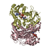
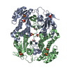
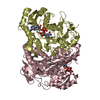
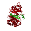
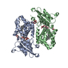
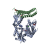
| Title | Crystal structure of a putative ketoacyl reductase (FabG4) from Mycobacterium tuberculosis H37Rv at 1.9 Angstrom resolution | ||||||
 Components Components | 3-oxoacyl-(Acyl-carrier-protein) reductase | ||||||
 Keywords Keywords | OXIDOREDUCTASE / Rossmann fold / Ketoreductase / High Molecular Weight FabG (HMwFabG) | ||||||
| Function / homology |  Function and homology information Function and homology informationlong-chain fatty-acyl-CoA metabolic process / short-chain fatty acid metabolic process / 3-oxoacyl-[acyl-carrier-protein] reductase / 3-oxoacyl-[acyl-carrier-protein] reductase (NADPH) activity / peptidoglycan-based cell wall / response to antibiotic / nucleotide binding / extracellular region / plasma membrane / cytosol Similarity search - Function | ||||||
| Biological species |   Mycobacterium tuberculosis (bacteria) Mycobacterium tuberculosis (bacteria) | ||||||
| Method |  X-RAY DIFFRACTION / MOLECULAR REPLACEMENT / Resolution: 1.88 Å X-RAY DIFFRACTION / MOLECULAR REPLACEMENT / Resolution: 1.88 Å | ||||||
 Authors Authors | Dutta, D. / Bhattacharyya, S. / Das, A.K. | ||||||
 Citation Citation | Journal: To be Published Title: Crystal structure of a putative ketoacyl reductase (FabG4) from Mycobacterium tuberculosis H37Rv at 1.9 Angstrom resolution Authors: Dutta, D. / Bhattacharyya, S. / Das, A.K. | ||||||
| History |
|
- Structure visualization
| Structure viewer | Molecule: MolmilJmol/JSmol |
|---|
- Downloads & links
-Download
| PDBx/mmCIF format | 3v1t.cif.gz | 311.6 KB | Display | PDBx/mmCIF format |
|---|---|---|---|---|
| PDB format | pdb3v1t.ent.gz | 249.9 KB | Display | PDB format |
| PDBx/mmJSON format | 3v1t.json.gz | Tree view | PDBx/mmJSON format | |
| Others |  Other downloads Other downloads |
-Validation report
| Arichive directory | https://data.pdbj.org/pub/pdb/validation_reports/v1/3v1tftp://data.pdbj.org/pub/pdb/validation_reports/v1/3v1t | HTTPS FTP |
|---|
-Related structure data
| Related structure data |  3q6iS S: Starting model for refinement |
|---|---|
| Similar structure data |


-Links
 PDBj
PDBj


- Assembly
| Deposited unit | 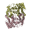
| ||||||||
|---|---|---|---|---|---|---|---|---|---|
| 1 |
| ||||||||
| Unit cell |
|
-Components
| #1: Protein | Mass: 47851.012 Da / Num. of mol.: 2 Source method: isolated from a genetically manipulated source Source: (gene. exp.) Mycobacterium tuberculosis (bacteria) / Strain: H37Rv / Gene: fabG-1, fabG4, MT0256, Rv0242c / Plasmid: pQE30 / Production host: References: UniProt: O53665, 3-oxoacyl-[acyl-carrier-protein] reductase #2: Water | ChemComp-HOH / |  Mass: 18.015 Da / Num. of mol.: 664 / Source method: isolated from a natural source / Formula: H2O Mass: 18.015 Da / Num. of mol.: 664 / Source method: isolated from a natural source / Formula: H2O |
|---|
-Experimental details
-Experiment
| Experiment | Method: X-RAY DIFFRACTION / Number of used crystals: 1 |
|---|
- Sample preparation
| Crystal | Density Matthews: 2.04 Å3/Da / Density % sol: 39.78 % / Mosaicity: 0 ° |
|---|---|
| Crystal grow | Temperature: 300 K / Method: vapor diffusion, hanging drop / pH: 6.5 Details: 45%(v/v) polypropylene glycol, 0.1M MES pH 6.5, VAPOR DIFFUSION, HANGING DROP, temperature 300K |
-Data collection
| Diffraction | Mean temperature: 103 K | ||||||||||||||||||||||||||||||||||||||||||||||||||||||||||||||||||||||||||||||||||||||||||||||||||||||||||||||||||||||||||||||||||||
|---|---|---|---|---|---|---|---|---|---|---|---|---|---|---|---|---|---|---|---|---|---|---|---|---|---|---|---|---|---|---|---|---|---|---|---|---|---|---|---|---|---|---|---|---|---|---|---|---|---|---|---|---|---|---|---|---|---|---|---|---|---|---|---|---|---|---|---|---|---|---|---|---|---|---|---|---|---|---|---|---|---|---|---|---|---|---|---|---|---|---|---|---|---|---|---|---|---|---|---|---|---|---|---|---|---|---|---|---|---|---|---|---|---|---|---|---|---|---|---|---|---|---|---|---|---|---|---|---|---|---|---|---|---|
| Diffraction source | Source: ROTATING ANODE / Type: RIGAKU MICROMAX-007 HF / Wavelength: 1.54 Å | ||||||||||||||||||||||||||||||||||||||||||||||||||||||||||||||||||||||||||||||||||||||||||||||||||||||||||||||||||||||||||||||||||||
| Detector | Type: RIGAKU RAXIS IV++ / Detector: IMAGE PLATE / Date: Dec 2, 2011 | ||||||||||||||||||||||||||||||||||||||||||||||||||||||||||||||||||||||||||||||||||||||||||||||||||||||||||||||||||||||||||||||||||||
| Radiation | Monochromator: MIRROR / Protocol: SINGLE WAVELENGTH / Monochromatic (M) / Laue (L): M / Scattering type: x-ray | ||||||||||||||||||||||||||||||||||||||||||||||||||||||||||||||||||||||||||||||||||||||||||||||||||||||||||||||||||||||||||||||||||||
| Radiation wavelength | Wavelength: 1.54 Å / Relative weight: 1 | ||||||||||||||||||||||||||||||||||||||||||||||||||||||||||||||||||||||||||||||||||||||||||||||||||||||||||||||||||||||||||||||||||||
| Reflection | Resolution: 1.876→66.392 Å / Num. all: 56389 / Num. obs: 56389 / % possible obs: 91 % / Redundancy: 1.9 % / Rsym value: 0.057 / Net I/σ(I): 8 | ||||||||||||||||||||||||||||||||||||||||||||||||||||||||||||||||||||||||||||||||||||||||||||||||||||||||||||||||||||||||||||||||||||
| Reflection shell | Diffraction-ID: 1
|
- Processing
| Software |
| |||||||||||||||||||||||||||||||||||||||||||||||||||||||||||||||||||||||||||
|---|---|---|---|---|---|---|---|---|---|---|---|---|---|---|---|---|---|---|---|---|---|---|---|---|---|---|---|---|---|---|---|---|---|---|---|---|---|---|---|---|---|---|---|---|---|---|---|---|---|---|---|---|---|---|---|---|---|---|---|---|---|---|---|---|---|---|---|---|---|---|---|---|---|---|---|---|
| Refinement | Method to determine structure: MOLECULAR REPLACEMENT Starting model: PDB ENTRY 3Q6I Resolution: 1.88→66.39 Å / Cor.coef. Fo:Fc: 0.956 / Cor.coef. Fo:Fc free: 0.914 / Occupancy max: 1 / Occupancy min: 1 / SU B: 8.623 / SU ML: 0.116 / Cross valid method: THROUGHOUT / σ(F): 0 / ESU R: 0.17 / ESU R Free: 0.172 / Stereochemistry target values: MAXIMUM LIKELIHOOD / Details: HYDROGENS HAVE BEEN ADDED IN THE RIDING POSITIONS
| |||||||||||||||||||||||||||||||||||||||||||||||||||||||||||||||||||||||||||
| Solvent computation | Ion probe radii: 0.8 Å / Shrinkage radii: 0.8 Å / VDW probe radii: 1.4 Å / Solvent model: MASK | |||||||||||||||||||||||||||||||||||||||||||||||||||||||||||||||||||||||||||
| Displacement parameters | Biso max: 117.49 Å2 / Biso mean: 26.298 Å2 / Biso min: 4.27 Å2
| |||||||||||||||||||||||||||||||||||||||||||||||||||||||||||||||||||||||||||
| Refinement step | Cycle: LAST / Resolution: 1.88→66.39 Å
| |||||||||||||||||||||||||||||||||||||||||||||||||||||||||||||||||||||||||||
| Refine LS restraints |
| |||||||||||||||||||||||||||||||||||||||||||||||||||||||||||||||||||||||||||
| LS refinement shell | Resolution: 1.876→1.925 Å / Total num. of bins used: 20
| |||||||||||||||||||||||||||||||||||||||||||||||||||||||||||||||||||||||||||
| Refinement TLS params. | Method: refined / Refine-ID: X-RAY DIFFRACTION
| |||||||||||||||||||||||||||||||||||||||||||||||||||||||||||||||||||||||||||
| Refinement TLS group |
|
