| 登録情報 | データベース: PDB / ID: 3v1o
|
|---|
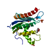
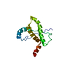
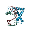
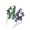
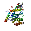
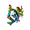
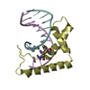
| タイトル | Crystal structures of the reverse transcriptase-associated ribonuclease H domain of xenotropic murine leukemia-virus related virus |
|---|
 要素 要素 | Reverse transcriptase/ribonuclease H p80 |
|---|
 キーワード キーワード | HYDROLASE / Reverse Transcription |
|---|
| 機能・相同性 |  機能・相同性情報 機能・相同性情報
ribonuclease H / 加水分解酵素; プロテアーゼ; ペプチド結合加水分解酵素; アスパラギン酸プロテアーゼ / virion assembly / DNA integration / viral genome integration into host DNA / RNA-directed DNA polymerase / establishment of integrated proviral latency / RNA-directed DNA polymerase activity / RNA-DNA hybrid ribonuclease activity / 転移酵素; リンを含む基を移すもの; 核酸を移すもの ...ribonuclease H / 加水分解酵素; プロテアーゼ; ペプチド結合加水分解酵素; アスパラギン酸プロテアーゼ / virion assembly / DNA integration / viral genome integration into host DNA / RNA-directed DNA polymerase / establishment of integrated proviral latency / RNA-directed DNA polymerase activity / RNA-DNA hybrid ribonuclease activity / 転移酵素; リンを含む基を移すもの; 核酸を移すもの / viral nucleocapsid / DNA recombination / DNA-directed DNA polymerase / structural constituent of virion / aspartic-type endopeptidase activity / 加水分解酵素; エステル加水分解酵素 / DNA-directed DNA polymerase activity / symbiont entry into host cell / host cell plasma membrane / proteolysis / DNA binding / RNA binding / zinc ion binding / membrane類似検索 - 分子機能 Gag-Pol polyprotein, Zinc-finger like domain / Murine leukemia virus integrase, C-terminal / Zinc-finger like, probable DNA-binding / Murine leukemia virus (MLV) integrase (IN) C-terminal domain / Gamma-retroviral matrix protein / Gag polyprotein, inner coat protein p12 / Core shell protein Gag P30 / Matrix protein (MA), p15 / Gag polyprotein, inner coat protein p12 / Gag P30 core shell protein ...Gag-Pol polyprotein, Zinc-finger like domain / Murine leukemia virus integrase, C-terminal / Zinc-finger like, probable DNA-binding / Murine leukemia virus (MLV) integrase (IN) C-terminal domain / Gamma-retroviral matrix protein / Gag polyprotein, inner coat protein p12 / Core shell protein Gag P30 / Matrix protein (MA), p15 / Gag polyprotein, inner coat protein p12 / Gag P30 core shell protein / Gamma-retroviral matrix domain superfamily / : / Reverse transcriptase/retrotransposon-derived protein, RNase H-like domain / RNase H-like domain found in reverse transcriptase / Ribonuclease H-like superfamily/Ribonuclease H / RNase H / Integrase core domain / Integrase, catalytic core / Integrase catalytic domain profile. / RNase H type-1 domain profile. / Ribonuclease H domain / Retropepsins / Retroviral aspartyl protease / Aspartyl protease, retroviral-type family profile. / Peptidase A2A, retrovirus, catalytic / Reverse transcriptase domain / Reverse transcriptase (RNA-dependent DNA polymerase) / Reverse transcriptase (RT) catalytic domain profile. / Retroviral matrix protein / Retrovirus capsid, N-terminal / zinc finger / Zinc knuckle / Zinc finger, CCHC-type superfamily / Zinc finger, CCHC-type / Zinc finger CCHC-type profile. / Nucleotidyltransferase; domain 5 / Aspartic peptidase, active site / Eukaryotic and viral aspartyl proteases active site. / Aspartic peptidase domain superfamily / Ribonuclease H superfamily / Ribonuclease H-like superfamily / Reverse transcriptase/Diguanylate cyclase domain / DNA/RNA polymerase superfamily / 2-Layer Sandwich / Alpha Beta類似検索 - ドメイン・相同性 |
|---|
| 生物種 |  Xenotropic MuLV-related virus VP35 (ウイルス) Xenotropic MuLV-related virus VP35 (ウイルス) |
|---|
| 手法 |  X線回折 / シンクロトロン / 分子置換 / 解像度: 1.876 Å X線回折 / シンクロトロン / 分子置換 / 解像度: 1.876 Å |
|---|
 データ登録者 データ登録者 | Zhou, D. / Wlodawer, A. |
|---|
 引用 引用 | ジャーナル: J.Struct.Biol. / 年: 2012
タイトル: Crystal structures of the reverse transcriptase-associated ribonuclease H domain of xenotropic murine leukemia-virus related virus.
著者: Zhou, D. / Chung, S. / Miller, M. / Le Grice, S.F. / Wlodawer, A. |
|---|
| 履歴 | | 登録 | 2011年12月9日 | 登録サイト: RCSB / 処理サイト: RCSB |
|---|
| 改定 1.0 | 2012年3月14日 | Provider: repository / タイプ: Initial release |
|---|
| 改定 1.1 | 2012年3月28日 | Group: Database references |
|---|
| 改定 1.2 | 2024年2月28日 | Group: Data collection / Database references / Derived calculations
カテゴリ: chem_comp_atom / chem_comp_bond ...chem_comp_atom / chem_comp_bond / database_2 / struct_ref_seq_dif / struct_site
Item: _database_2.pdbx_DOI / _database_2.pdbx_database_accession ..._database_2.pdbx_DOI / _database_2.pdbx_database_accession / _struct_ref_seq_dif.details / _struct_site.pdbx_auth_asym_id / _struct_site.pdbx_auth_comp_id / _struct_site.pdbx_auth_seq_id |
|---|
|
|---|
 ムービー
ムービー コントローラー


 データを開く
 基本情報
基本情報 構造の表示
構造の表示 ダウンロードとリンク
 その他のダウンロード
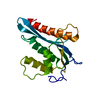


 PDBj
PDBj
 集合体
集合体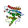


 分子量: 96.063 Da / 分子数: 4 / 由来タイプ: 合成 / 式: SO4
 分子量: 118.174 Da / 分子数: 1 / 由来タイプ: 合成 / 式: C6H14O2 / コメント: 沈殿剤*YM
 分子量: 92.094 Da / 分子数: 1 / 由来タイプ: 合成 / 式: C3H8O3
 分子量: 18.015 Da / 分子数: 71 / 由来タイプ: 天然 / 式: H2O
 試料調製
試料調製 / ビームライン: 22-ID / 波長: 1 Å
 解析
解析